. 2015 Sep 11:5:14007.
doi: 10.1038/srep14007.

Detection of Langat virus by TaqMan real-time one-step qRT-PCR method

Affiliations

Detection of Langat virus by TaqMan real-time one-step qRT-PCR method

Siti Fatimah Muhd Radzi et al. Sci Rep. .

Abstract

Langat virus (LGTV), one of the members of the tick-borne encephalitis virus (TBEV) complex, was firstly isolated from Ixodes granulatus ticks in Malaysia. However, the prevalence of LGTV in ticks in the region remains unknown. Surveillance for LGTV is therefore important and thus a tool for specific detection of LGTV is needed. In the present study, we developed a real-time quantitative reverse-transcription-polymerase chain reaction (qRT-PCR) for rapid detection of LGTV. Our findings showed that the developed qRT-PCR could detect LGTV at a titre as low as 0.1 FFU/ml. The detection limit of the qRT-PCR assay at 95% probability was 0.28 FFU/ml as determined by probit analysis (p ≤ 0.05). Besides, the designed primers and probe did not amplify ORF of the E genes for some closely related and more pathogenic viruses including TBEV, Louping ill virus, Omsk hemorrhagic fever virus (OHFV), Alkhurma virus (ALKV), Kyasanur Forest Disease virus (KFDV) and Powassan virus (POWV) which showed the acceptable specificity of the developed assay. The sensitivity of the developed method also has been confirmed by determining the LGTV in infected tick cell line as well as LGTV- spiked tick tissues.


Conflict of interest statement

The authors declare no competing financial interests.

Figures
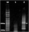
Figure 1
Figure 1. Confirmation of RT-PCR amplification for TBEV RNA.
Lane M, 100 bp plus ladder; lane 1, negative control; lane 2, TBEV RNA. A RT-PCR product of the expected size (532 bp) is visible in lane 2.
Figure 2
Figure 2. Map of LGTV-specific qRT-PCR primers and TaqMan probe in alignment with the viral E gene sequences of the LGTV related flaviviruses.
The ORF for the E gene of Louping ill virus, Omsk hemorrhagic fever virus, Alkhumra virus, Kyasanur Forest disease virus, and Powassan virus have been aligned with the related ORF in LGTV genome using Clustal X 2.0 software. The arrows indicate the orientation of primers in 5′ to 3′ direction.
Figure 3
Figure 3. Detection limit of the qRT-PCR for detection of LGTV in nuclease-free water.
The probit regression curve was obtained from nine replicates of LGTV RNA at seven dilutions (100, 50, 10, 5, 1, 0.5 and 0.1 FFU/ml).
Figure 4
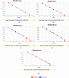
Figure 4. Standard curves for qRT-PCRs.
(a) Standard curve of qRT-PCR for detection of LGTV. The amplification efficiency (Eff%) was 101.988 and coefficient of determination (R2) was 1. (b) Standard curve for specificity of LGTV qRT-PCR against five closely related viruses. The Eff% of qRT-PCR was 118.189 and the R2 was 0.998. The LGTV qRT-PCR assay didn’t amplify any of five closely related viruses while the negative control of the qRT-PCR was undetermined. (c) Standard curve for specificity of LGTV qRT-PCR against in vitro transcribed TBEV RNA. The Eff% of qRT-PCR was 117.976 and the R2 was 0.999. There was no in vitro transcribed TBEV RNA detected. (d) Standard curve of qRT-PCR for detection of LGTV RNA in spiked homogenized tick tissues. The Eff% of qRT-PCR was 108.91and the R2 was 0.998. LGTV RNA detected in spiked sample with LGTV at 212.24 FFU/ml and the other sample was spiked with TBEV and LGTV together at 239.11 FFU/ml (Blue boxes). (e) Standard curve of qRT-PCR for detection of LGTV RNA in IDE8 cells. The Eff% of qRT-PCR was 135.054 and the R2 was 0.995. LGTV RNA in IDE8 cells was detected at 406,268 FFU/ml (2 days poi) and 776,412 FFU/ml (4 days poi) (Blue boxes).

References

    1. Chambers T. J., Hahn C. S., Gailer R. & Rice C. M. Flavivirus genome organization,expression, and replication. Annu Rev Microbiol. 44, 649–688 (1990). - PubMed
    1. Smith C. G. A virus resembling Russian spring–summer encephalitis virus from an Ixodid tick in Malaya. Nature 178, 581–582 (1956). - PubMed
    1. Bancroft W. H., Scott R., Snitbhan R., Weaver R. Jr & Gould D. J. Isolation of Langat virus from Haemaphysalis papuanaThorell in Thailand. Am J Trop Med Hyg. 25, 500–504 (1976). - PubMed
    1. Seamer J. & Randles W. The course of Langat virus infection in mice. Br J Exp Pathol. 48, 403 (1967). - PMC - PubMed
    1. Webb H., Wight D., Platt G. & Smith C. Langat virus encephalitis in mice I. The effect of the administration of specific antiserum. J Hyg (Lond) 66, 343–354 (1968). - PMC - PubMed

Publication types

LinkOut - more resources