. 2015 Sep 11;10(9):e0137745.
doi: 10.1371/journal.pone.0137745. eCollection 2015.

The Protein Tyrosine Phosphatase Rptpζ Suppresses Osteosarcoma Development in Trp53-Heterozygous Mice

Affiliations

The Protein Tyrosine Phosphatase Rptpζ Suppresses Osteosarcoma Development in Trp53-Heterozygous Mice

Christina Baldauf et al. PLoS One. .

Abstract

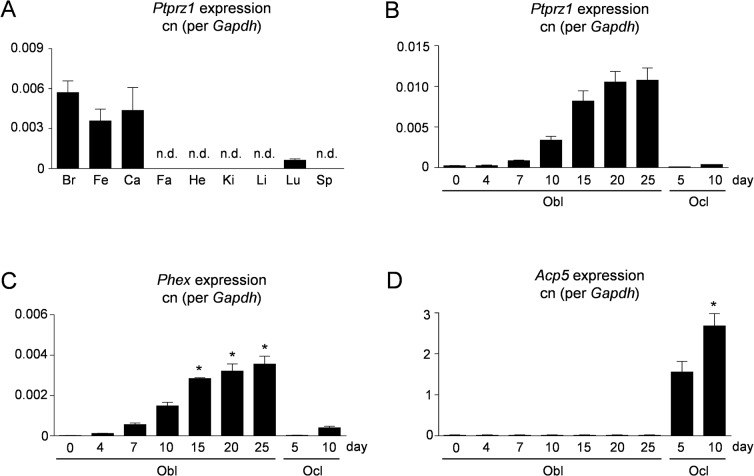
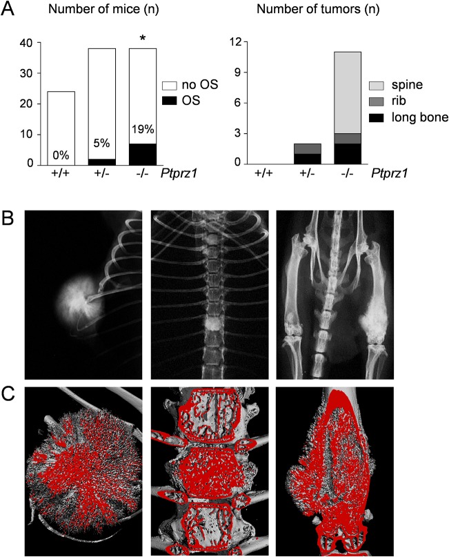
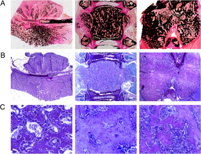
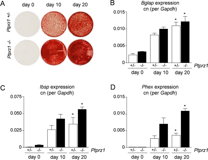
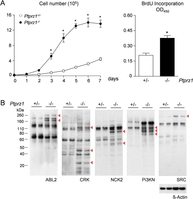
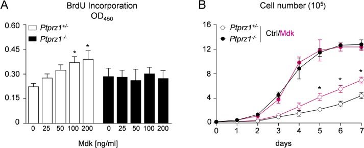
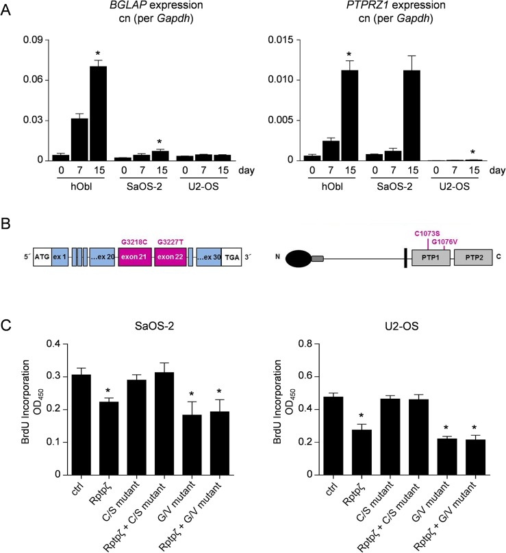
Osteosarcoma (OS), a highly aggressive primary bone tumor, belongs to the most common solid tumors in growing children. Since specific molecular targets for OS treatment remain to be identified, surgical resection combined with multimodal (neo-)adjuvant chemotherapy is still the only way to help respective individuals. We have previously identified the protein tyrosine phosphatase Rptpζ as a marker of terminally differentiated osteoblasts, which negatively regulates their proliferation in vitro. Here we have addressed the question if Rptpζ can function as a tumor suppressor protein inhibiting OS development in vivo. We therefore analyzed the skeletal phenotype of mice lacking Ptprz1, the gene encoding Rptpζ on a tumor-prone genetic background, i.e. Trp53-heterozygosity. By screening a large number of 52 week old Trp53-heterozygous mice by contact radiography we found that Ptprz1-deficiency significantly enhanced OS development with 19% of the mice being affected. The tumors in Ptprz1-deficient Trp53-heterozygous mice were present in different locations (spine, long bones, ribs), and their OS nature was confirmed by undecalcified histology. Likewise, cell lines derived from the tumors were able to undergo osteogenic differentiation ex vivo. A comparison between Ptprz1-heterozygous and Ptprz1-deficient cultures further revealed that the latter ones displayed increased proliferation, a higher abundance of tyrosine-phosphorylated proteins and resistance towards the influence of the growth factor Midkine. Our findings underscore the relevance of Rptpζ as an attenuator of proliferation in differentiated osteoblasts and raise the possibility that activating Rptpζ-dependent signaling could specifically target osteoblastic tumor cells.


Conflict of interest statement

Competing Interests: The authors have declared that no competing interests exist.

Figures

Fig 1
Fig 1. Ptprz1 expression by differentiated osteoblasts.
(A) qRT-PCR monitoring Ptprz1 expression in brain (Br), femur (Fe), calvaria (Ca), fat (Fa), heart (He), kidney (Ki), liver (Li), lung (Lu) and spleen (Sp). (B) qRT-PCR monitoring Ptprz1 expression in primary osteoblasts (Obl) or osteoclasts (Ocl) at different stages of differentiation. Bars represent mean ± SD (n = 3). (C) qRT-PCR monitoring expression of the osteocyte marker Phex. (D) qRT-PCR monitoring expression of the osteoclast marker Acp5 (encoding TRAP). Values represent copy number (cn) relative to Gadph. Bars represent mean ± SD (n = 3). Asterisks indicate statistical significance vs. Obl day 0 (p<0.05, Kruskal-Wallis followed by Dunn’s post-test).
Fig 2
Fig 2. Skeletal tumors in Ptprz1-deficient Trp53-heterozygous mice.
(A) OS development assessed by screening of 12 month old Trp53-heterozygous mice with the indicated Ptprz1 genotypes. The left panel shows the number of analyzed mice (white bars) and the percentage of mice with OS (black bars). The asterisk indicates statistical significance vs. Trp53 +/- /Ptprz1 +/+ (p<0.05, two-tailed Fishers`s exact test). The right panel shows the total number of tumors and their location. (B) Representative contact Xrays from Ptprz1-deficient Trp53-heterozygous mice with OS in the three different locations. (C) μCT images from the same tumors.
Fig 3
Fig 3. OS nature of skeletal tumors in Ptprz1-deficient Trp53-heterozygous mice.
(A) Von Kossa/Van Gieson staining of undecalficied sections confirms that the tumors contain mineralized matrix (stained black). (B) Toluidine blue staining demonstrating dark blue staining of cartilage areas and light blue staining of the tumors. (C) Higher magnification images reveal that the tumors represented bony tissue with osteocytes embedded into the mineralized matrix.
Fig 4
Fig 4. Osteogenic differentiation of OS cell lines.
(A) Alizarin red staining of cells derived from Trp53-heterozygous mice with either one Ptprz1 allele (+/-) or with Ptprz1-deficiency (-/-) reveals that both cell lines are able to form a mineralized matrix after 10 and 20 days of differentiation induced by ascorbic acid and ß-glycerophosphate. (B-D) qRT-PCR expression analysis shows that the differentiation is accompanied by increased expression of Bglap (encoding Osteocalcin), Ibsp (encoding Bone sialoprotein) and Phex. Values represent copy number (cn) relative to Gadph. Bars represent mean ± SD (n = 3). Asterisks indicate significant differences towards day 0 of corresponding genotype (p<0.05, Kruskal-Wallis followed by Dunn’s post-test).
Fig 5
Fig 5. Proliferation and tyrosine phosphorylation in OS cell lines.
(A) Proliferative capacity of tumor cells derived from Trp53-heterozygous mice with either one Ptprz1 allele (+/-) or with Ptprz1-deficiency (-/-). The growth curves (left) and the BrdU incorporation assays (right) demonstrate increased proliferation in the cases of Ptprz1-deficiency. Bars represent mean ± SD (n≥3). Asterisks indicate significant differences between the two genotypes (p<0.05, two-way ANOVA followed by Bonferroni’s post-test (left panel) or two-tailed Student’s t-test (right panel)). (B) SH2 profiling with different SH2 domains reveals differences in tyrosine phosphorylation of specific proteins (indicated by arrowheads). Re-probing of stripped membranes with anti ß-actin mAb served as control for equal loading. Analyses were performed in duplicate with cell extracts harvested at different cell densities.
Fig 6
Fig 6. Effects of Mdk on proliferation of OS cell lines.
(A) BrdU incorporation assays with the indicated OS cell lines performed in the presence of Mdk at different concentrations. Bars represent mean ± SD (n = 8). Asterisks indicate significant differences towards controls without Mdk (p<0.05, one-way ANOVA followed by Dunnett’s post-test). (B) Growth curves of OS cell lines in the presence or absence of Mdk (100 ng/ml). Asterisks indicate significant differences towards controls of Ptprz1 +/- cells without Mdk (p<0.05, two-way ANOVA followed by Bonferroni’s post-test).
Fig 7
Fig 7. Functional analysis of Rptpζ mutations.
(A) qRT-PCR monitoring expression of BGLAP (left) and PTPRZ1 (right) in human primary osteoblasts (hObl), SaOS-2 or U2-OS cells at different stages of differentiation. Values represent copy number (cn) relative to GAPDH. Bars represent mean ± SD (n = 3). Asterisks indicate statistical significance vs. day 0 (p<0.05, Kruskal-Wallis followed by Dunn’s post-test). (B) Schematic presentation of the PTPRZ1 gene and Rptpζ protein showing the location of the two mutations that have been introduced into a Rptpζ expression plasmid. (C) BrdU incorporation assay with SaOS-2 and U2-OS cells after transfection of wildtype and/or mutant Rptpζ expression plasmids as indicated. Bars represent mean ± SD (n = 6). Asterisks indicate significant differences towards cells transfected with empty vector (ctrl) (p<0.05, one-way ANOVA followed by Dunnett’s post-test).

References

    1. Kuijjer ML, Hogendoorn PC, Cleton-Jansen AM. (2013) Genome-wide analyses on high-grade osteosarcoma: making sense of a genomically most unstable tumor. Int J Cancer 133: 2512–2521. 10.1002/ijc.28124 - DOI - PubMed
    1. Lietman SA, Joyce MJ. (2010) Bone sarcomas: Overview of management, with a focus on surgical treatment considerations. Cleve Clin J Med 77: S8–S12. 10.3949/ccjm.77.s1.02 - DOI - PubMed
    1. Pakos EE, Nearchou AD, Grimer RJ, Koumoullis HD, Abudu A, Bramer JA, et al. (2009) Prognostic factors and outcomes for osteosarcoma: an international collaboration. Eur J Cancer 45: 2367–2375. 10.1016/j.ejca.2009.03.005 - DOI - PubMed
    1. Bacci G, Bertoni F, Longhi A, Ferrari S, Forni C, Biagini R, et al. (2003) Neoadjuvant chemotherapy for high-grade central osteosarcoma of the extremity. Histologic response to preoperative chemotherapy correlates with histologic subtype of the tumor. Cancer 97: 3068–3075. - PubMed
    1. Allison DC, Carney SC, Ahlmann ER, Hendifar A, Chawla S, Fedenko A, et al. (2011) A meta-analysis of osteosarcoma outcomes in the modern medical era. Sarcoma 2012: 704872. - PMC - PubMed

Publication types

MeSH terms

Substances