

Segmentation of the thalamus based on BOLD frequencies affected in temporal lobe epilepsy
- PMID: 26360535
- PMCID: PMC4626388
- DOI: 10.1111/epi.13186
Segmentation of the thalamus based on BOLD frequencies affected in temporal lobe epilepsy
Abstract
Objective: Temporal lobe epilepsy is associated with functional changes throughout the brain, particularly including a putative seizure propagation network involving the hippocampus, insula, and thalamus. We identified a specified frequency range where functional connectivity in this network was related to duration of disease. Then, to identify specific thalamic nuclei involved in seizure propagation, we determined the subregions of the thalamus that have increased resting functional oscillations in this frequency range.
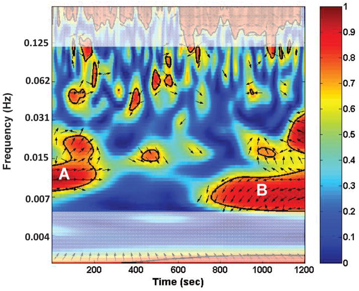
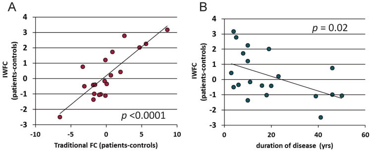
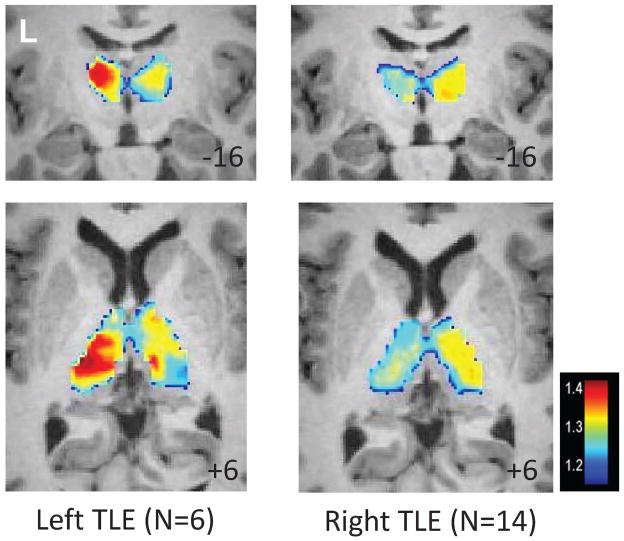
Methods: Resting-state functional magnetic resonance imaging (fMRI) was acquired from 20 patients with unilateral temporal lobe epilepsy (TLE; 14 right and 6 left) and 20 healthy controls who were each age and gender matched to a specific patient. Wavelet-based fMRI connectivity mapping across the network was computed at each frequency to determine those frequencies where connectivity significantly decreases with duration of disease consistent with impairment due to repeated seizures. The voxel-wise power of the spontaneous blood oxygenation fluctuations of this frequency band was computed in the thalamus of each subject.
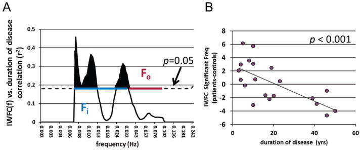
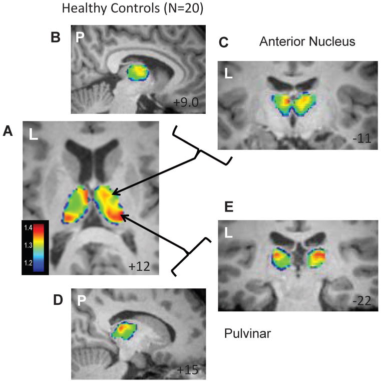
Results: Functional connectivity was impaired in the proposed seizure propagation network over a specific range (0.0067-0.013 Hz and 0.024-0.032 Hz) of blood oxygenation oscillations. Increased power in this frequency band (<0.032 Hz) was detected bilaterally in the pulvinar and anterior nucleus of the thalamus of healthy controls, and was increased over the ipsilateral thalamus compared to the contralateral thalamus in TLE.
Significance: This study identified frequencies of impaired connectivity in a TLE seizure propagation network and used them to localize the anterior nucleus and pulvinar of the thalamus as subregions most susceptible to TLE seizures. Further examinations of these frequencies in healthy and TLE subjects may provide unique information relating to the mechanism of seizure propagation and potential treatment using electrical stimulation.
Keywords: Functional neuroimaging; Temporal lobe epilepsy; Thalamus; fMRI.
Wiley Periodicals, Inc. © 2015 International League Against Epilepsy.
Conflict of interest statement
None of the authors has any conflict of interest to disclose. We confirm that we have read the Journal’s position on issues involved in ethical publication and affirm that this report is consistent with those guidelines.
Figures
References
-
- Biswal B, Yetkin FZ, Haughton VM, et al. Functional connectivity in the motor cortex of resting human brain using echo-planar MRI. Magn Reson Med. 1995;34:537–41. - PubMed
-
- Blauwblomme T, David O, Minotti L, et al. Prognostic value of insular lobe involvement in temporal lobe epilepsy: A stereoelectroencephalographic study. Epilepsia. 2013;54:1658–1667. - PubMed
-
- Isnard J, Guenot M, Ostrowsky K, et al. The role of the insular cortex in temporal lobe epilepsy. Ann Neurol. 2000;48:614–23. - PubMed
Publication types
MeSH terms
Grants and funding
LinkOut - more resources
Full Text Sources
Other Literature Sources
Medical
