

Evidence for Functionally Relevant Encounter Complexes in Nitrogenase Catalysis
- PMID: 26360912
- PMCID: PMC4809638
- DOI: 10.1021/jacs.5b08310
Evidence for Functionally Relevant Encounter Complexes in Nitrogenase Catalysis
Abstract
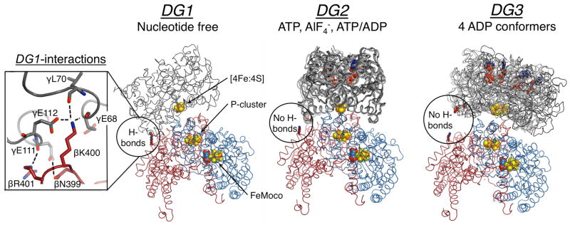
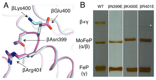
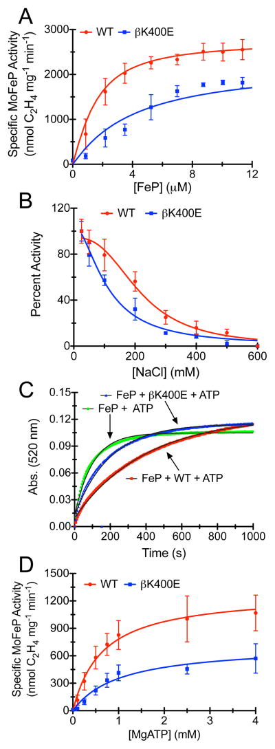
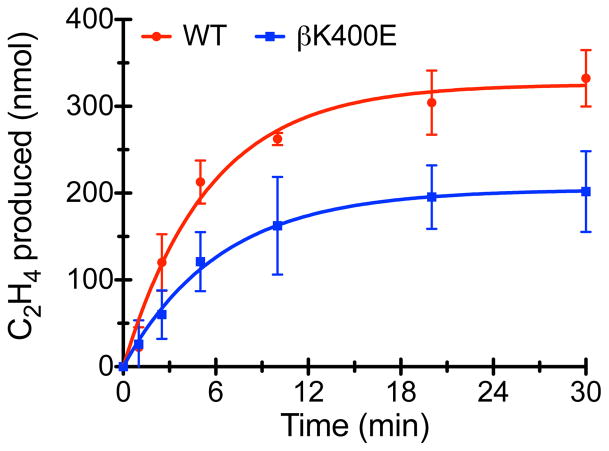
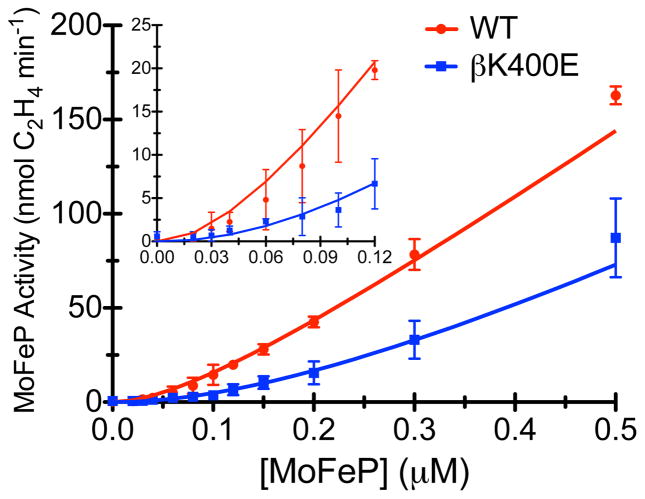
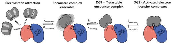
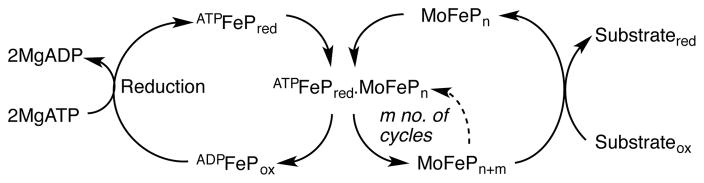
Nitrogenase is the only enzyme that can convert atmospheric dinitrogen (N2) into biologically usable ammonia (NH3). To achieve this multielectron redox process, the nitrogenase component proteins, MoFe-protein (MoFeP) and Fe-protein (FeP), repeatedly associate and dissociate in an ATP-dependent manner, where one electron is transferred from FeP to MoFeP per association. Here, we provide experimental evidence that encounter complexes between FeP and MoFeP play a functional role in nitrogenase catalysis. The encounter complexes are stabilized by electrostatic interactions involving a positively charged patch on the β-subunit of MoFeP. Three single mutations (βAsn399Glu, βLys400Glu, and βArg401Glu) in this patch were generated in Azotobacter vinelandii MoFeP. All of the resulting variants displayed decreases in specific catalytic activity, with the βK400E mutation showing the largest effect. As simulated by the Thorneley-Lowe kinetic scheme, this single mutation lowered the rate constant for FeP-MoFeP association 5-fold. We also found that the βK400E mutation did not affect the coupling of ATP hydrolysis with electron transfer (ET) between FeP and MoFeP. These data suggest a mechanism where FeP initially forms encounter complexes on the MoFeP β-subunit surface en route to the ATP-activated, ET-competent complex over the αβ-interface.
Figures
Similar articles
-
A voltammetric study of nitrogenase MoFe-protein using low-potential electron transfer mediators.Bioelectrochemistry. 2024 Feb;155:108575. doi: 10.1016/j.bioelechem.2023.108575. Epub 2023 Sep 17. Bioelectrochemistry. 2024. PMID: 37738860
-
Docking of nitrogenase iron- and molybdenum-iron proteins for electron transfer and MgATP hydrolysis: the role of arginine 140 and lysine 143 of the Azotobacter vinelandii iron protein.Protein Sci. 1994 Nov;3(11):2073-81. doi: 10.1002/pro.5560031120. Protein Sci. 1994. PMID: 7703853 Free PMC article.
-
Formation of a tight 1:1 complex of Clostridium pasteurianum Fe protein-Azotobacter vinelandii MoFe protein: evidence for long-range interactions between the Fe protein binding sites during catalytic hydrogen evolution.Biochemistry. 2000 Sep 19;39(37):11434-40. doi: 10.1021/bi0002939. Biochemistry. 2000. PMID: 10985789
-
Nitrogenase structure and function: a biochemical-genetic perspective.Annu Rev Microbiol. 1995;49:335-66. doi: 10.1146/annurev.mi.49.100195.002003. Annu Rev Microbiol. 1995. PMID: 8561464 Review.
-
Electron Transfer in Nitrogenase.Chem Rev. 2020 Jun 24;120(12):5158-5193. doi: 10.1021/acs.chemrev.9b00663. Epub 2020 Jan 30. Chem Rev. 2020. PMID: 31999100 Free PMC article. Review.
Cited by
-
Mechanical coupling in the nitrogenase complex.PLoS Comput Biol. 2021 Mar 4;17(3):e1008719. doi: 10.1371/journal.pcbi.1008719. eCollection 2021 Mar. PLoS Comput Biol. 2021. PMID: 33661889 Free PMC article.
-
Crystal structure of VnfH, the iron protein component of vanadium nitrogenase.J Biol Inorg Chem. 2018 Oct;23(7):1049-1056. doi: 10.1007/s00775-018-1602-4. Epub 2018 Aug 23. J Biol Inorg Chem. 2018. PMID: 30141094
-
Conformationally Gated Electron Transfer in Nitrogenase. Isolation, Purification, and Characterization of Nitrogenase From Gluconacetobacter diazotrophicus.Methods Enzymol. 2018;599:355-386. doi: 10.1016/bs.mie.2017.09.007. Epub 2017 Dec 6. Methods Enzymol. 2018. PMID: 29746246 Free PMC article.
-
Preparation of oxygen-sensitive proteins for high-resolution cryoEM structure determination using blot-free vitrification.Nat Commun. 2025 Apr 14;16(1):3528. doi: 10.1038/s41467-025-58243-1. Nat Commun. 2025. PMID: 40229244 Free PMC article.
-
Statistical analysis of PN clusters in Mo/VFe protein crystals using a bond valence method toward their electronic structures.RSC Adv. 2022 Feb 11;12(9):5214-5224. doi: 10.1039/d1ra08507g. eCollection 2022 Feb 10. RSC Adv. 2022. PMID: 35425536 Free PMC article.
References
Publication types
MeSH terms
Substances
Grants and funding
LinkOut - more resources
Full Text Sources
Other Literature Sources
Research Materials
