. 2015 Sep 11;10(9):e0137423.
doi: 10.1371/journal.pone.0137423. eCollection 2015.

Indoleamine 2,3-Dioxygenase Is Involved in the Inflammation Response of Corneal Epithelial Cells to Aspergillus fumigatus Infections

Affiliations

Indoleamine 2,3-Dioxygenase Is Involved in the Inflammation Response of Corneal Epithelial Cells to Aspergillus fumigatus Infections

Nan Jiang et al. PLoS One. .

Abstract

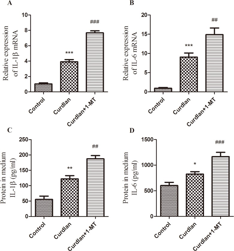
Indoleamine 2,3-dioxygenase (IDO), which is mainly expressed in activated dendritic cells, is known as a regulator of immune responses. However, the role of IDO in immune responses against fungal corneal infection has not been investigated. To evaluate the regulatory mechanisms of IDO in fungal inflammation, we resorted to human corneal epithelial cells (HCECs), known as the first barrier of cornea against pathogenic microorganisms. We found that IDO was significantly up-regulated in corneal epithelium infected with Aspergillus fumigatus (A. fumigatus) and HCECs incubated with spores of A. fumigatus. Furthermore, IDO inhibitor (1-methyltryptophan, 1-MT) enhanced inflammatory cytokines IL-1β and IL-6 expression which were up-regulated by A. fumigatus spores infection. Dectin-1, as one of the important C-type lectin receptors, can identify β-glucan, and mediate fungal innate immune responses. In the present study, pre-treatment with curdlan, a Dectin-1 agonist, further enhanced IDO expression compared with A. fumigatus stimulation. While laminarin, the Dectin-1 specific inhibitor, partially inhibited IDO expression stimulated by A. fumigatus. Further studies demonstrated inhibition of IDO activity amplified the expressions of inflammatory cytokines IL-1β and IL-6 induced by activation of Dectin-1. These results suggested that IDO was involved in the immune responses of fungal keratitis. The activation of Dectin-1 may contribute to A. fumigatus spores-induced up-regulation of IDO.


Conflict of interest statement

Competing Interests: The authors have declared that no competing interests.

Figures

Fig 1
Fig 1. Immunofluorescence localization of IDO in normal and infected human corneas.
Serial sections of normal and A. fumigatus-infected corneas were immunostained using IDO antibody and FITC-conjugated anti-mouse second antibody (for IDO, green), followed by counterstaining with DAPI (blue). Note that in normal cornea arrayed in the first row, no immunoreactivity was detected in corneal epithelium, while strong green fluorescence in A. fumigatus-infected corneal epithelium by immunofluorescent assays. Enlarged images clearly displayed IDO staining was primarily localized in the cytoplasm of corneal epithelial cells in the infected cornea. Scale bar in immunofluorescence images: 50μm.
Fig 2
Fig 2. Expression of IDO mRNA in human corneal epithelium with fungal infection.
qRT-PCR was performed to identify relative expression of IDO mRNA in corneal epithelium. Normal corneal tissue remainings after corneal transplantation were considered controls. Corneas with fungal infections were classified into three grades (mild, moderate and severe) according to keratomycosis severity. Fungal infection increased IDO mRNA expression in the human corneal epithelium and the expression correlated with keratomycosis severity. The data were represented as the mean ± standard deviation of four independent experiments. (**p < 0.01, ***p < 0.001 compared with control; ## p < 0.01, compared with mild grade group; $ $ $ p < 0.001 compared with moderate grade group).
Fig 3
Fig 3. IDO expression in cultured telomerase-immortalized HCECs infected with A. fumigatus.
Relative expression of IDO mRNA was detected by qRT-PCR 4, 10, 16 and 24 h after incubation with A. fumigatus spores at three different concentrations (5 × 10⁶ /ml, 5 × 10⁷ /ml and 5 × 10⁸ /ml). IDO expression increased by 4- to 5-fold with a peak level at 10 h after exposure to inactivated A. fumigatus spores (5 × 10⁷ /ml).
Fig 4
Fig 4. IL-1β and IL-6 expressions after blocking of IDO in cultured telomerase-immortalized HCECs infected with A. fumigatus.
qRT-PCR and ELISA data demonstrated that incubation with A. fumigatus led to up-regulation of IL-1β (A, C) and IL-6 (B, D) compared with controls. However, 1-MT-induced blockade of IDO further significantly enhanced the expressions of the above inflammatory cytokines at 10 h post-infection. The data were represented as mean ± standard deviation of six independent experiments. (*p < 0.05, ***p < 0.001 compared with control; # p < 0.05, ## p < 0.01, ### p < 0.001 compared with A. fumigatus treatment group).
Fig 5
Fig 5. 1-MT treatment enhanced inflammatory cytokine production in corneas of mice infected with A. fumigatus.
C57BL/6 mice were inoculated with 2 μl of 1 × 10⁵/μl spores. (A) The corneas were monitored and taken photograps under a slit lamp, and (B) evaluated with the scoring system. Disease score is shown as mean ± standard deviation. After 1-MT treatment, the IL-1β mRNA and protein levels were significantly increased (C, D) at 3 days after infection in 1-MT treated mice compared with PBS-treated mice. In addition, both IL-6 mRNA (E) and IL-6 protein (F) were significantly increased at 3 days after infection. In addition, the IL-1β proteins were increased at 5 days after infection in 1-MT treated mice. (*p < 0.05, **p < 0.001 compared with PBS treated mice).
Fig 6
Fig 6. Induction of IDO by microbial ligands in HCECs.
Levels of IDO mRNA at 10 h after stimulation by 200 μg/ml curdlan or 10μg/ml of Pam3CSK4, LPS were evaluated by qRT-PCR. Data were represented as mean ± standard deviation of three independent experiments. (*p< 0.05, **p < 0.01).
Fig 7
Fig 7. The effect of Dectin-1 on IDO expressions in cultured HCECs infected with A. fumigatus.
The HCECs were pre-treated with curdlan or laminarin, and were exposed to inactivated A. fumigatus spores for 10 h. The cultures were subjected to qRT-PCR to measure IDO mRNA expressions (A). The cultures treated for 24 h were used to evaluate IDO protein expressions in HCECs by western blot (B). The data are represented as mean ± standard deviation of four independent experiments. (*p < 0.05, **p < 0.01).
Fig 8
Fig 8. The effects of 1-MT on curdlan mediated IL-1β and IL-6 expressions in HCECs.
The HCECs were pre-treated by curdlan and/or 1-MT. The cultures treated for 10 h were subjected to qRT-PCR for mRNA measurement (A, B), the cultures treated for 24 h were used to evaluate protein in the medium of 6-well by ELISA (C, D). The data were represented as mean ± standard deviation of six independent experiments. (*p < 0.05, **p < 0.01, ***p < 0.001 compared with control; ## p < 0.01, ### p < 0.001 compared with curdlan treatment group).

References

    1. Kredics L, Narendran V, Shobana CS, Vágvölgyi C, Manikandan P, Indo-Hungarian Fungal Keratitis Working Group. Filamentous fungal infections of the cornea: a global overview of epidemiology and drug sensitivity. Mycoses. 2015; 58(4):243–260. 10.1111/myc.12306 - DOI - PubMed
    1. Sharma Y, Jain S, Jayachandran. Keratomycosis: Etiology, Risk Factors and Differential Diagnosis- A Mini Review on Trichophyton spp. J Clin Diagn Res. 2014;8: DD01–02. - PMC - PubMed
    1. Xuguang S, Zhixin W, Zhiqun W, Shiyun L, Ran L. Ocular fungal isolates and antifungal susceptibility in northern China. Am J Ophthalmol. 2007;143: 131–133. - PubMed
    1. Xie L, Zhong W, Shi W, Sun S. Spectrum of fungal keratitis in north China. Ophthalmology. 2006;113: 1943–1948. - PubMed
    1. Wang L, Sun S, Jing Y, Han L, Zhang H, Yue J. Spectrum of fungal keratitis in central China. Clin Experiment Ophthalmol. 2009;37: 763–771. 10.1111/j.1442-9071.2009.02155.x - DOI - PubMed

Publication types

MeSH terms