Review
. 2015 Sep;47(10):727-34.
doi: 10.1055/s-0035-1559633. Epub 2015 Sep 11.

Graves' Disease Mechanisms: The Role of Stimulating, Blocking, and Cleavage Region TSH Receptor Antibodies

Affiliations
Review

Graves' Disease Mechanisms: The Role of Stimulating, Blocking, and Cleavage Region TSH Receptor Antibodies

S A Morshed et al. Horm Metab Res. 2015 Sep.

Abstract

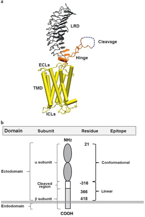
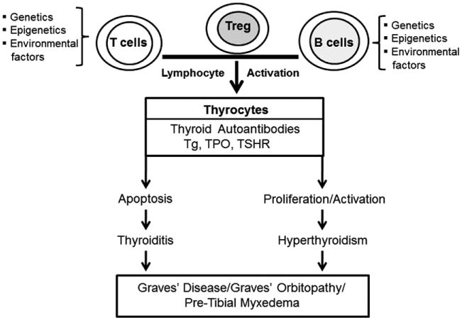
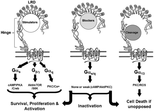
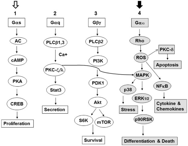
The immunologic processes involved in Graves' disease (GD) have one unique characteristic--the autoantibodies to the TSH receptor (TSHR)--which have both linear and conformational epitopes. Three types of TSHR antibodies (stimulating, blocking, and cleavage) with different functional capabilities have been described in GD patients, which induce different signaling effects varying from thyroid cell proliferation to thyroid cell death. The establishment of animal models of GD by TSHR antibody transfer or by immunization with TSHR antigen has confirmed its pathogenic role and, therefore, GD is the result of a breakdown in TSHR tolerance. Here we review some of the characteristics of TSHR antibodies with a special emphasis on new developments in our understanding of what were previously called "neutral" antibodies and which we now characterize as autoantibodies to the "cleavage" region of the TSHR ectodomain.


Figures

Fig. 1
Fig. 1
a A computer generated model of the TSHR based on the crystal structure of the ectodomain with the 7 transmembrane domain structure derived from the rhodopsin receptor crystal. The large ectodomain consists of 9 leucine-rich repeats (LRRs), which form a characteristic “horse shoe” shaped structure with a concave inner surface which harbors the major ligand and TSHR-Ab binding regions. The cleavage region and unique 50 amino acids cleaved region in the ectodomain, of unknown structure, is shown in gray and is a unique characteristic of the TSHR. b A schematic model of the TSHR to illustrate the multiple domains, subunits and epitope distributions containing different amino acid residues. TSHR stimulating and blocking antibodies are directed to the conformational epitopes whereas cleavage antibodies are directed at linear epitopes, mainly targeting the cleavage region.
Fig. 2
Fig. 2
A simplified outline of the factors contributing to the development of Graves' disease on a background of thyroiditis.
Fig. 3
Fig. 3
The 3 different varieties of TSHR-Abs and their signal transduction pathways and functional consequences.
Fig. 4
Fig. 4
Although still highly complex, this is a simplified TSHR signaling model for the major G-protein activation pathways. In FRTL-5 thyrocytes, TSHR-stimulating antibodies and TSH itself activate the major signaling cascades numbered 1, 2, and 3. In contrast, cleavage TSHR-Abs activate mainly cascade #4 in grey (with the closed arrow at the top). Signaling cascade #4 appears to be exclusive to the cleavage antibodies and induces Rho, ROS and other down-stream signaling effectors such as NFκB and PKC-δ. In this proposed model system, Gαs activates cAMP/PKA/CREB via adenyl cyclase activation and Gαq induces PKC/Stat3 via activation of PLCβ1.3 kinases with Ca + [40]. Similarly, Gβγ activates PI3 kinase and downstream molecules PDK1/Akt/mTOR/S6k1 via PLCβ2 kinase. PKC (curved arrow) once activated can induce different MAPKs including ERK1/2/p38/p90RSK.
Fig. 5
Fig. 5
An outline of the proposed mechanisms involved in cleavage TSHR-Ab induced apoptosis (in FRTL-5) thyrocytes. While stimulating antibodies induce thyrocyte cell survival and proliferation via cAMP/PKA/CREB and Akt/mTOR/S6K, cleavage antibodies would do the opposite if unopposed and result in cell death via ROS induction. Since cleavage antibodies are unable to activate Gαs, Gαq, and Gβγ (unpublished), they may activate Gα13 and its downstream effectors Rho/ROS/PKC-δ leading to apoptosis via activation of the Cyt-C/Bax signaling cascade (unpublished).
Fig. 6
Fig. 6
Immunohistochemistry and live imaging of stress markers in thyrocytes. Panel a Immunohistochemical detection of stress-induced proteins. Rabbit polyclonal primary antibody was used to detect Mn-SOD in C-TSHR-mAb (IC8 1 μg/ml) treated (24 h) FRTL-5 thyrocytes. The C-TSHR-mAb also induced heat shock proteins (HSP) 70 and 60 as detected by mAbs against each protein. HSP70 and 60 are normally localized within the endoplasmic reticulum and mitochondria, respectively. Such proteins were not induced by an isotype control mAb (see insets). Panel b Live imaging of ROS in thyrocytes using 3 independent dyes. Cells were treated for 24 h with C-TSHR-mAb (IC8 1 μg/ml). Representative images of both D123 and H2DCFDA showed diffuse staining throughout the cell cytoplasm. Some perinuclear condensations were also documented [16]. The distribution of staining patterns paralleled mitochondrial ROS (mROS). Scale bar on images corresponds to 20 micron. Nucleoli are indicated as N.

References

    1. Bahn RS, Dutton CM, Natt N, Joba W, Spitzweg C, Heufelder AE. Thyrotropin receptor expression in Graves' orbital adipose/connective tissues: potential autoantigen in Graves' ophthalmopathy. J Clin Endocrinol Metab. 1998;83:998–1002. - PubMed
    1. Chazenbalk GD, Pichurin P, Chen CR, Latrofa F, Johnstone AP, McLachlan SM, Rapoport B. Thyroid-stimulating autoantibodies in Graves disease preferentially recognize the free A subunit, not the thyrotropin holoreceptor. J Clin Invest. 2002;110:209–217. - PMC - PubMed
    1. Kajita Y, Rickards CR, Buckland PR, Howells RD, Rees SB. Analysis of thyrotropin receptors by photoaffinity labelling. Orientation of receptor subunits in the cell membrane. Biochem J. 1985;227:413–420. - PMC - PubMed
    1. Loosfelt H, Pichon C, Jolivet A, Misrahi M, Caillou B, Jamous M, Vannier B, Milgrom E. Two-subunit structure of the human thyrotropin receptor. Proc Natl Acad Sci USA. 1992;89:3765–3769. - PMC - PubMed
    1. Vlase H, Davies TF. Insights into the molecular mechanisms of the autoimmune thyroid diseases. In: Eisenbarth GS, editor. Endocrine and Organ Specific Autoimmunity. CA: R. G. Landes Co; 1999. pp. 98–132.

Publication types