. 2015 Sep 11;10(9):e0136620.
doi: 10.1371/journal.pone.0136620. eCollection 2015.

Functional Role of the Disulfide Isomerase ERp57 in Axonal Regeneration

Affiliations

Functional Role of the Disulfide Isomerase ERp57 in Axonal Regeneration

Valentina Castillo et al. PLoS One. .

Erratum in

Abstract

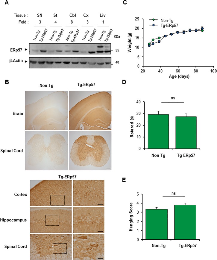
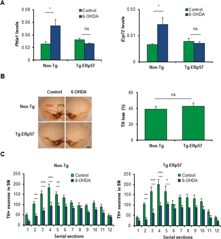
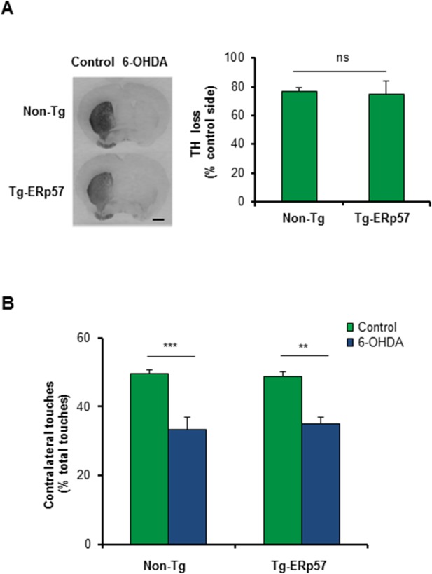
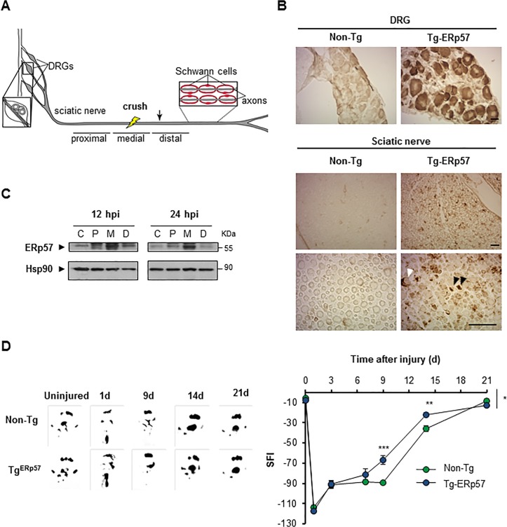
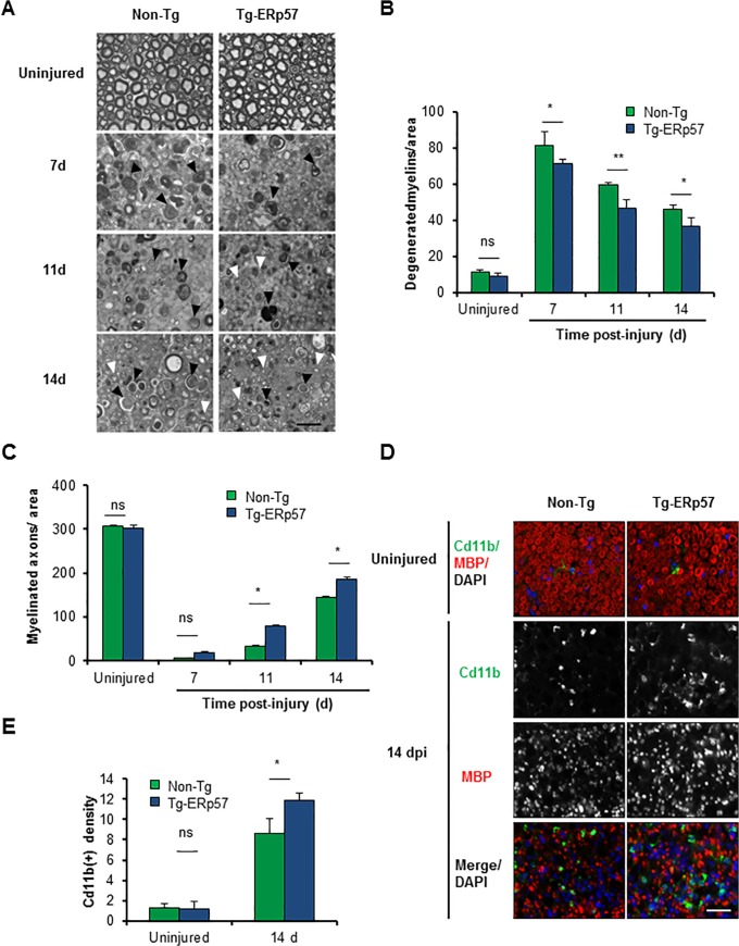
ERp57 (also known as grp58 and PDIA3) is a protein disulfide isomerase that catalyzes disulfide bonds formation of glycoproteins as part of the calnexin and calreticulin cycle. ERp57 is markedly upregulated in most common neurodegenerative diseases downstream of the endoplasmic reticulum (ER) stress response. Despite accumulating correlative evidence supporting a neuroprotective role of ERp57, the contribution of this foldase to the physiology of the nervous system remains unknown. Here we developed a transgenic mouse model that overexpresses ERp57 in the nervous system under the control of the prion promoter. We analyzed the susceptibility of ERp57 transgenic mice to undergo neurodegeneration. Unexpectedly, ERp57 overexpression did not affect dopaminergic neuron loss and striatal denervation after injection of a Parkinson's disease-inducing neurotoxin. In sharp contrast, ERp57 transgenic animals presented enhanced locomotor recovery after mechanical injury to the sciatic nerve. These protective effects were associated with enhanced myelin removal, macrophage infiltration and axonal regeneration. Our results suggest that ERp57 specifically contributes to peripheral nerve regeneration, whereas its activity is dispensable for the survival of a specific neuronal population of the central nervous system. These results demonstrate for the first time a functional role of a component of the ER proteostasis network in peripheral nerve regeneration.


Conflict of interest statement

Competing Interests: The authors have declared that no competing interests exist.

Figures

Fig 1
Fig 1. Generation of an ERp57 transgenic mouse model.
(A) ERp57 protein levels were analyzed in nervous system tissue of a neuronal specific ERp57 transgenic (Tg-ERp57) mice and littermate control non-transgenic (Non-Tg) animals using Western blot. The following tissue was analyzed: substantia nigra (SN), striatum (St), cerebellum (Cbl), cortex (Cx) and liver (Liv). β-Actin was used as a loading control. In the upper panel, quantification of the relative ERp57 levels is presented normalized to Non-Tg animals. (B) Immunohistochemistry anti-FLAG was performed in nervous system tissue of Tg-ERp57 mice and littermate control non-transgenic (Non-Tg). The following tissue was analyzed: cortex, hippocampus, and spinal cord ventral horn. At the right panels, magnification of indicated areas is shown. Scale bar: 200 μm for lower magnifications and 50 μm (left) and 25 μm (right) for higher magnification. (C) Analysis of body weight in Tg-ERp57 and Non-Tg animals over time. (D) Rotarod performance was measured every week starting at 50 days of age in animals presented in C. (E) Hanging test was performed every week starting at approximately at 50 days of age as described in material and methods. In C-D mean ± SEM is presented of each group. Tg-ERp57 (n = 15) and Non-Tg (n = 18) animals. Student’s unpaired t-test was performed to calculate statistical significance (*: p < 0.05; n.s.: non-significant).
Fig 2
Fig 2. Effects of ERp57 overexpression on the survival of dopaminergic neurons after exposure to 6-OHDA.
(A) Pdia1 and Erp72 mRNA levels were determined in dissected SNpc of Non-Tg and Tg-ERp57 mice 7 d after 6-OHDA injection. Injected and non-injected sides were analyzed using total cDNA and real-time PCR (n = 3 per group). Data is presented as mean and SEM. (B) Non-Tg and Tg-ERp57 mice were injected with 8 μg of 6-OHDA in the right striatum, and after 7d dopaminergic neurons (TH+) were quantified by anti-TH immunohistochemistry. Total content of TH-positive somas was measured in midbrain sections covering the entire SN, in the non-injected (control) and injected (6-OHDA) side, for the indicated genotype (n = 8, Non-Tg; n = 5, Tg-ERp57). Scale bar: 200 μm. (C) Histograms show the number of TH-positive neurons of injected and non-injected sides in 25 μm midbrain serial sections separated by 100 μm and covering the entire SNpc. The numbers of serial sections indicate the orientation from anterior to posterior. Statistical analysis was performed using Mann-Whitney test for all quantifications except for (C) where two-way ANOVA was used followed by Bonferroni posttest (*: p < 0.05; **: p < 0.01; ***: p < 0.001. n.s.: not significant.).
Fig 3
Fig 3. ERp57 overexpression does not affect 6-OHDA-induced striatal denervation or motor control.
(A) Immunohistochemistry analysis was performed in striatal sections to quantify 6-OHDA–induced denervation in both injected (6-OHDA) and non-injected (control) sides. Scale bar: 1 mm. Right panel: The integrated density of pixel intensity was calculated from images of anti-TH immunohistochemistry covering the entire striatum and expressed as a percentage of TH loss relative to the control side (n = 8, Non-Tg; n = 5, Tg-ERp57). Scale bar: 200 μm. (B) Cylinder test was performed to evaluate spontaneous motor changes associated with dopamine depletion in the striatum of 6-OHDA injected mice. The cylinder test consist in to put the animals in a glass and record the number times the mouse touch the glass wall with each forepaw. Non-Tg (n = 10) and Tg-ERp57 (n = 8). Data is presented as mean and SEM. Statistical analyses were performed using Mann-Whitney test (A) and the Student t test (B). (*: p < 0.05; **: p < 0.01; ***: p < 0.001. n.s.: not significant.)
Fig 4
Fig 4. Overexpression of ERp57 in transgenic mice enhances locomotor recovery after peripheral nerve injury.
(A) Scheme of a peripheral nerve: Sciatic nerve is formed by the spinal nerves of the dorsal root ganglia (DRG) from the L3, L4 and L5 vertebrae. DRG contains somas of sensory neurons and axons from motoneurons located in the ventral horn of spinal cord. Schwann cells surround peripheral axons to form myelinated and unmyelinated fibers. Nerves were damaged by mechanical crush (yellow bolt) and a 5 mm region from the injury region and adjacent proximal and distal regions were removed for biochemical analysis. A 3 mm region distal to the injury was removed for histological analysis (arrow). (B) Immunohistochemistry anti-FLAG was performed in DRG and sciatic nerve (Sci) of Tg-ERp57 mice and littermate control non-transgenic (Non-Tg). Black and white arrowheads points immunoreactive axons and Schwann cells, respectively. Scale bar: 40 μm. (C) Wild-type mice were damaged in the right sciatic nerve and ERp57 expression was evaluated at 12 and 24 h post injury (hpi) in proximal, P, medial, M and distal, D region. Contralateral sham-operated nerves, C were used as controls. Images were obtained from the analysis of the same gel and membrane. Hsp90 expression was used as loading control. (D) Non-Tg and Tg-ERp57 mice (n = 7 animals per group) were injured and the locomotor performance was analyzed using the sciatic nerve functional index (SFI). (Left panel) SFI was measured for 21 days after injury. (Right panel) Representative footprints of the damaged hind limb of Non-Tg and Tg-ERp57 mice at 0 (uninjured), 1, 9, 14 and 21 days post-injury. Data are shown as mean ± S.E.M. * p < 0.05, ** p < 0.01, *** p < 0.001. For SFI analysis data were analyzed by two-way ANOVA followed by Bonferroni post test.
Fig 5
Fig 5. Overexpression of ERp57 in transgenic mice increases axonal regeneration after peripheral nerve injury.
(A) Non-Tg and Tg-ERp57 mice were damaged and sciatic nerves were extracted at 7, 11 and 14 days post-injury (dpi). Transversal semi-thin sections were obtained from the distal region and remyelinated axons (white arrows) and degenerated myelins (black arrows) were analyzed. (B) Quantification of remyelinated axons and (C) degenerated myelin density was measured at 7, 11 and 14 days post-injury. (D) Tg- ERp57 and Non-Tg sciatic nerves were processed for immunofluorescence in uninjured conditions and at 14 dpi. Sciatic nerves were analyzed for MBP (red), Cd11b (green) and nuclei were stained using DAPI (blue). (E) The staining density for Cd11b was quantified 14 days after injury (right panel). Data is presented as mean ± S.E.M. * p < 0.05, ** p < 0.01, *** p < 0.001. For histological analysis, statistical differences were obtained using a student’s t-test (n = 3 animals per group). Scale bar: 20 μm.

References

    1. Soto C. Unfolding the role of protein misfolding in neurodegenerative diseases. Nature reviews Neuroscience. 2003;4(1):49–60. - PubMed
    1. Hetz C, Mollereau B. Disturbance of endoplasmic reticulum proteostasis in neurodegenerative diseases. Nature reviews Neuroscience. 2014;15(4):233–49. 10.1038/nrn3689 - DOI - PubMed
    1. Balch WE, Morimoto RI, Dillin A, Kelly JW. Adapting proteostasis for disease intervention. Science. 2008;319(5865):916–9. 10.1126/science.1141448 - DOI - PubMed
    1. Hetz C, Martinon F, Rodriguez D, Glimcher LH. The unfolded protein response: integrating stress signals through the stress sensor IRE1alpha. Physiological reviews. 2011;91(4):1219–43. 10.1152/physrev.00001.2011 - DOI - PubMed
    1. Walter P, Ron D. The unfolded protein response: from stress pathway to homeostatic regulation. Science. 2011;334(6059):1081–6. 10.1126/science.1209038 - DOI - PubMed

Publication types

MeSH terms