

Hepatic hemangioma -review-
Abstract
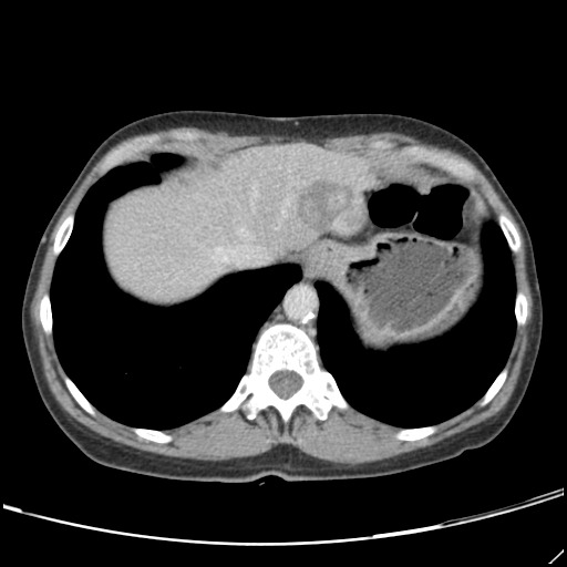
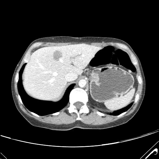
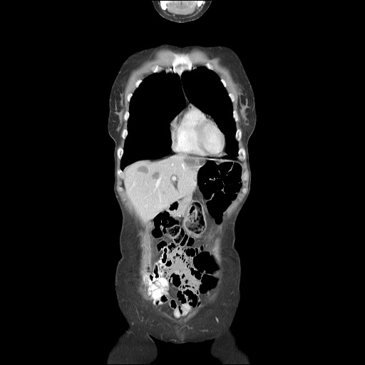
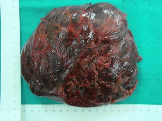
Hepatic hemangiomas are benign tumors of the liver consisting of clusters of blood-filled cavities, lined by endothelial cells, fed by the hepatic artery. The vast majority of HH are asymptomatic, most often being discovered incidentally during imaging investigations for various unrelated pathologies. Typical hemangiomas, the so-called capillary hemangiomas, range from a few mm to 3 cm, do not increase in size over time and therefore are unlikely to generate future symptomatology. Small (mm-3 cm) and medium (3 cm-10 cm) hemangiomas are well-defined lesions, requiring no active treatment beside regular follow-ups. However, the so-called giant liver hemangiomas, of up to 10 cm (most commonly) and even 20+ cm in size (according to occasional reports) can, and usually will develop symptoms and complications that require prompt surgical intervention or other kind of therapy. HH belong to the class of hepatic "incidentalomas", so-called because they are diagnosed incidentally, on imaging studies performed as routine examinations or for other reasons than the evaluation of a possible liver mass. Less than half of HH present with overt clinical symptoms, consisting, most often, of upper abdominal pain (this is usually the case for large lesions, which cause the distension of Glisson's capsule). Hepatic hemangiomas require a careful diagnosis to differentiate from other focal hepatic lesions, co-occurring diagnoses are also possible.
Keywords: computer tomography (CT); contrast-enhanced ultrasonography (CEUS); conventional ultrasonography (US); hepatic hemangioma (HH); ultrasonography contrast agent (UCA).
Figures


References
-
- Popescu I, et al. Chirurgia ficatului. 2004;1:141-142–231-232.
-
- Sechel G, Fleancu A, Rogozea L. Aspecte atipice imagistice ale hemangioamelor hepatice. J.M.B. :49–55.
Publication types
MeSH terms
LinkOut - more resources
Full Text Sources
Medical