

Data-driven optimized flip angle selection for T1 estimation from spoiled gradient echo acquisitions
- PMID: 26361720
- PMCID: PMC4788991
- DOI: 10.1002/mrm.25920
Data-driven optimized flip angle selection for T1 estimation from spoiled gradient echo acquisitions
Abstract
Purpose: Define criteria for selection of optimal flip angle sets for T1 estimation and evaluate effects on T1 mapping.
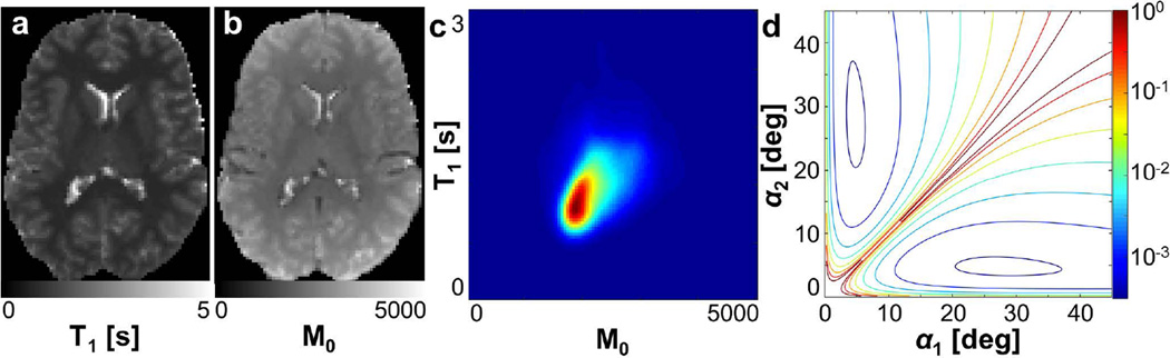
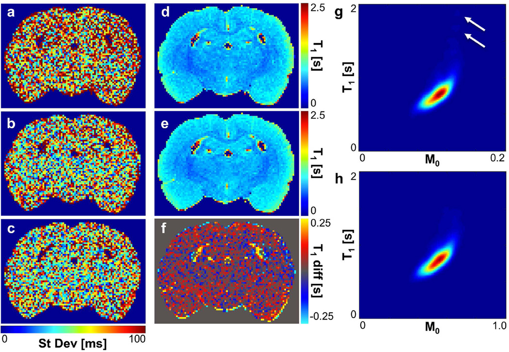
Theory and methods: Flip angle sets for spoiled gradient echo-based T1 mapping were selected by minimizing T1 estimate variance weighted by the joint density of M0 and T1 in an initial acquisition. The effect of optimized flip angle selection on T1 estimate error was measured using simulations and experimental data in the human and rat brain.
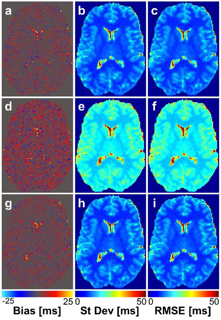
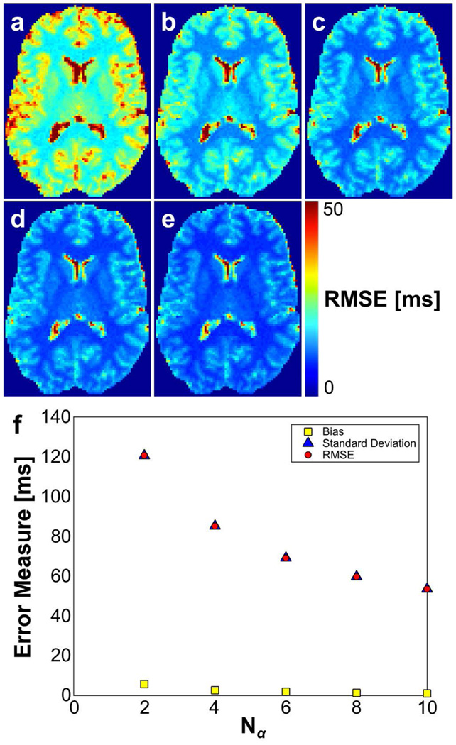
Results: For two-point acquisitions, optimized angle sets were similar to those proposed by other groups and, therefore, performed similarly. For multipoint acquisitions, optimal angle sets for T1 mapping in the brain consisted of a repetition of two angles. Implementation of optimal angles reduced T1 estimate variance by 30-40% compared with a multipoint acquisition using a range of angles. Performance of the optimal angle set was equivalent to that of a repetition of the two-angle set selected using criteria proposed by other researchers.
Conclusion: Repetition of two carefully selected flip angles notably improves the precision of resulting T1 estimates compared with acquisitions using a range of flip angles. This work provides a flexible and widely applicable optimization method of particular use for those who repeatedly perform T1 estimation. Magn Reson Med 76:792-802, 2016. © 2015 Wiley Periodicals, Inc.
Keywords: SPGR; T1 mapping; error propagation; flip angle selection.
© 2015 Wiley Periodicals, Inc.
Figures
References
-
- Lutti A, Dick F, Sereno MI, Weiskopf N. Using high-resolution quantitative mapping of R1 as an index of cortical myelination. Neuroimage. 2014;93:176–188. - PubMed
-
- Baudrexel S, Nurnberger L, Rub U, Seifried C, Klein JC, Deller T, Steinmetz H, Deichmann R, Hilker R. Quantitative mapping of T1 and T2* discloses nigral and brainstem pathology in early Parkinson's disease. Neuroimage. 2010;51:512–520. - PubMed
-
- Stikov N, Boudreau M, Levesque IR, Tardif CL, Barral JK, Pike GB. On the accuracy of T1 mapping: searching for common ground. Magn Reson Med. 2015;73:514–522. - PubMed
-
- Fram EK, Herfkens RJ, Johnson GA, Glover GH, Karis JP, Shimakawa A, Perkins TG, Pelc NJ. Rapid calculation of T1 using variable flip angle gradient refocused imaging. Magn Reson Imaging. 1987;5:201–208. - PubMed
Publication types
MeSH terms
Grants and funding
LinkOut - more resources
Full Text Sources
Other Literature Sources
Medical
