. 2015 Dec 1;24(23):6588-602.
doi: 10.1093/hmg/ddv363. Epub 2015 Sep 11.

Myofibrillar disruption and RNA-binding protein aggregation in a mouse model of limb-girdle muscular dystrophy 1D

Affiliations

Myofibrillar disruption and RNA-binding protein aggregation in a mouse model of limb-girdle muscular dystrophy 1D

Rocio Bengoechea et al. Hum Mol Genet. .

Abstract

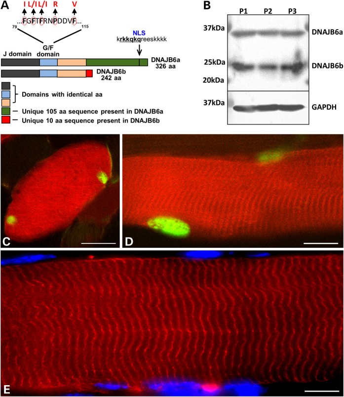
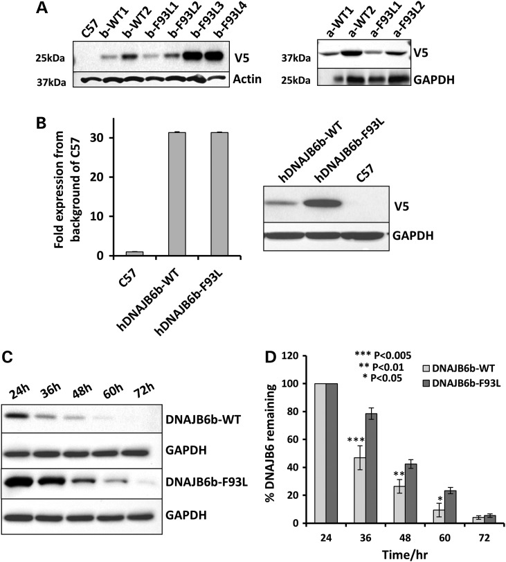
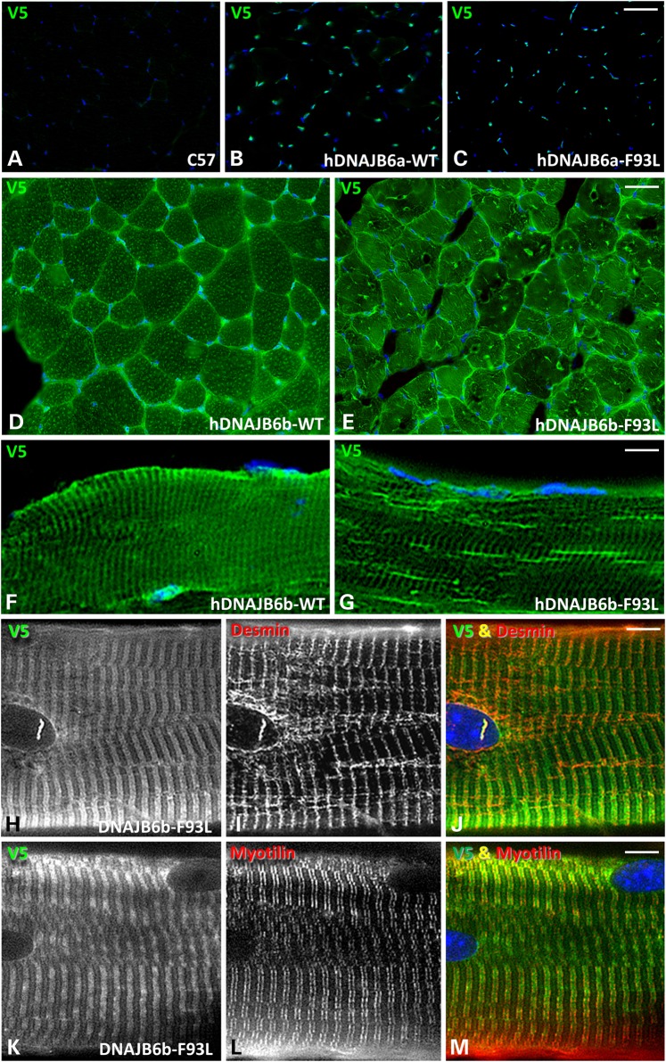
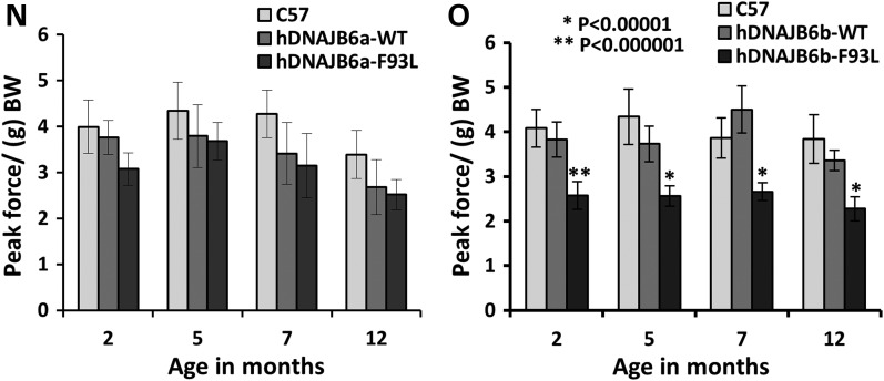
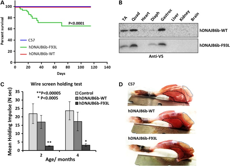
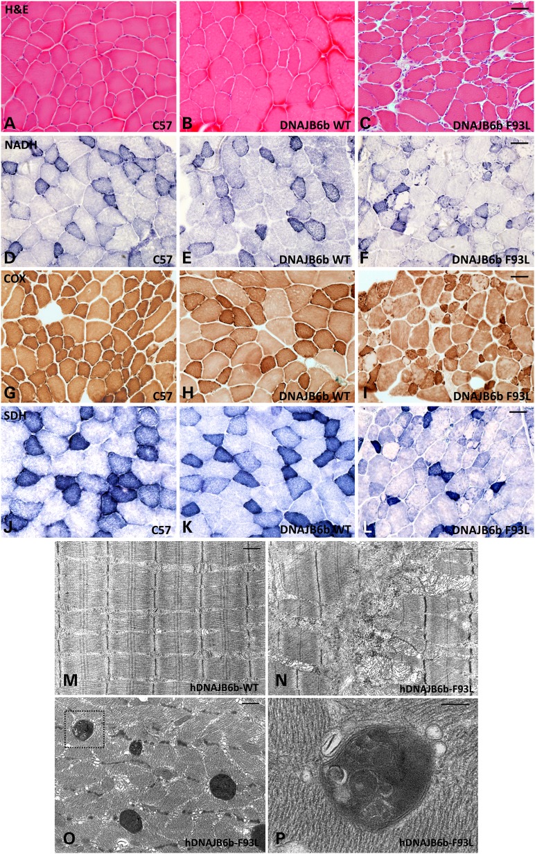
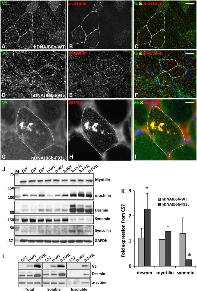
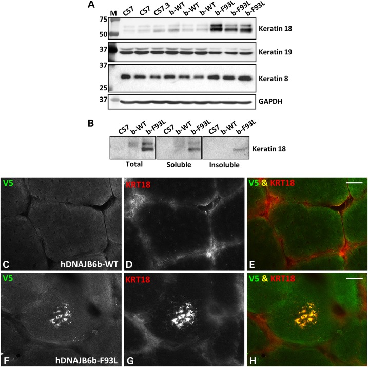
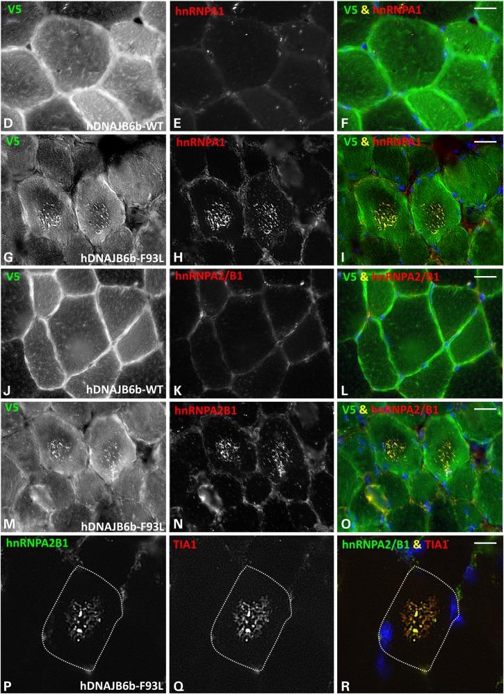
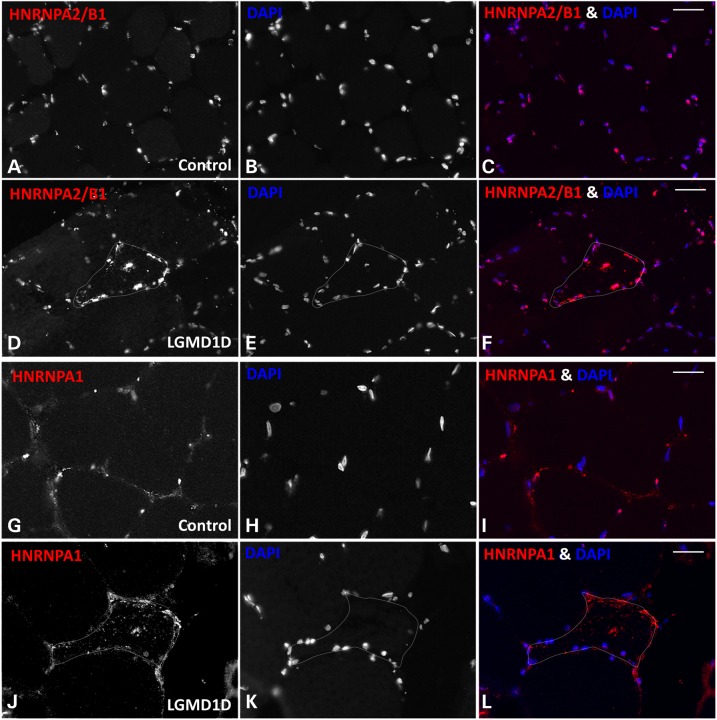
Limb-girdle muscular dystrophy type 1D (LGMD1D) is caused by dominantly inherited missense mutations in DNAJB6, an Hsp40 co-chaperone. LGMD1D muscle has rimmed vacuoles and inclusion bodies containing DNAJB6, Z-disc proteins and TDP-43. DNAJB6 is expressed as two isoforms; DNAJB6a and DNAJB6b. Both isoforms contain LGMD1D mutant residues and are expressed in human muscle. To identify which mutant isoform confers disease pathogenesis and generate a mouse model of LGMD1D, we evaluated DNAJB6 expression and localization in skeletal muscle as well as generating DNAJB6 isoform specific expressing transgenic mice. DNAJB6a localized to myonuclei while DNAJB6b was sarcoplasmic. LGMD1D mutations in DNAJB6a or DNAJB6b did not alter this localization in mouse muscle. Transgenic mice expressing the LGMD1D mutant, F93L, in DNAJB6b under a muscle-specific promoter became weak, had early lethality and developed muscle pathology consistent with myopathy after 2 months; whereas mice expressing the same F93L mutation in DNAJB6a or overexpressing DNAJB6a or DNAJB6b wild-type transgenes remained unaffected after 1 year. DNAJB6b localized to the Z-disc and DNAJB6b-F93L expressing mouse muscle had myofibrillar disorganization and desmin inclusions. Consistent with DNAJB6 dysfunction, keratin 8/18, a DNAJB6 client also accumulated in DNAJB6b-F93L expressing mouse muscle. The RNA-binding proteins hnRNPA1 and hnRNPA2/B1 accumulated and co-localized with DNAJB6 at sarcoplasmic stress granules suggesting that these proteins maybe novel DNAJB6b clients. Similarly, hnRNPA1 and hnRNPA2/B1 formed sarcoplasmic aggregates in patients with LGMD1D. Our data support that LGMD1D mutations in DNAJB6 disrupt its sarcoplasmic function suggesting a role for DNAJB6b in Z-disc organization and stress granule kinetics.


Figures

Figure 1.
Figure 1.
DNAJB6a and DNAJB6b expression in skeletal muscle. (A) Schematic of DNAJB6a and DNAJB6b isoforms. Note that all five LGMD1D mutant residues reside within the G/F domain that is common to both isoforms. (B) DNAJB6 immunoblot from three different human skeletal muscle lysates showing a DNAJB6a and DNAJB6b isoform. GAPDH is loading control. (CE) Mouse tibialis anterior muscle was electroporated with constructs expressing both mCherry-DNAJB6b and GFP-DNAJB6a (C and D) or mCherry-DNAJB6b alone (E). DNAJB6b (red) is sarcoplasmic and associates with sarcomeric structures whereas DNAJB6a (green) is exclusively myonuclear. Bar (C and D), 20 µm and Bar (E), 10 µm.
Figure 2.
Figure 2.
LGMD1D mutant DNAJB6b is more stable than DNAJB6b-WT in vivo. (A) Immunoblot of skeletal muscle lysates from control (C57), V5-hDNAJB6b-WT (b-WT), V5-hDNAJB6b-F93L (b-F93L), V5-hDNAJB6a-WT (a-WT) and V5-hDNAJB6a-F93L (a-F93L) expressing mice using a V5 antibody. Actin and GAPDH are loading controls. (B) Quantitative PCR expression matched V5-hDNAJB6b-WT and V5-hDNAJB6b-F93L lines and corresponding V5 immunoblot from the same lysate. Note that hDNAJBb-F93L is more stable. (C) Tetracycline inducible isogenic 293 cell lines expressing V5-DNAJB6b-WT or V5-DNAJB6b-F93L were induced for 24 h and then tetracycline was removed and lysates were immunoblotted for V5 and GAPDH at the indicated times. (D) Graphical representation of the results in C from three independent experiments.
Figure 3.
Figure 3.
DNAJB6b-F93L transgenic develop muscle weakness. (AC) Frozen sections of tibialis anterior from 4-month old control (C57), hDNAJB6a-WT or hDNAJB6a-F93L immunostained with V5 (green) and DAPI (blue). hDNAJB6a is localized to myonuclei. Bar, 100 µm. (DG) Frozen sections of tibialis anterior from 4-month old hDNAJB6b-WT and hDNAJBb-F93L expressing mice using an V5 antibody. Note that hDNAJB6b is localized to the sarcomere. Bar (D and E), 100 µm and Bar (FG), 20 µm. (HM) Teased fibers from V5-hDNAJB6b-F93L mice co-stained with V5 and desmin or myotilin. Note that hDNAJB6b localizes to the Z-disc. Bar, 10 µm. (N) Forelimb grip strength measurements from littermate control (C57), hDNAJB6a-WT and hDNAJB6a-F93L mice at 2, 5, 7 and 12 months of age. (O) Forelimb grip strength measurements from littermate control (C57), hDNAJB6b-WT and hDNAJB6b-F93L mice at 2, 5, 7 and 12 months of age. Note that hDNAJB6b-F93L mice develop persistent weakness at 2 months of age.
Figure 3.
Figure 3.
DNAJB6b-F93L transgenic develop muscle weakness. (AC) Frozen sections of tibialis anterior from 4-month old control (C57), hDNAJB6a-WT or hDNAJB6a-F93L immunostained with V5 (green) and DAPI (blue). hDNAJB6a is localized to myonuclei. Bar, 100 µm. (DG) Frozen sections of tibialis anterior from 4-month old hDNAJB6b-WT and hDNAJBb-F93L expressing mice using an V5 antibody. Note that hDNAJB6b is localized to the sarcomere. Bar (D and E), 100 µm and Bar (FG), 20 µm. (HM) Teased fibers from V5-hDNAJB6b-F93L mice co-stained with V5 and desmin or myotilin. Note that hDNAJB6b localizes to the Z-disc. Bar, 10 µm. (N) Forelimb grip strength measurements from littermate control (C57), hDNAJB6a-WT and hDNAJB6a-F93L mice at 2, 5, 7 and 12 months of age. (O) Forelimb grip strength measurements from littermate control (C57), hDNAJB6b-WT and hDNAJB6b-F93L mice at 2, 5, 7 and 12 months of age. Note that hDNAJB6b-F93L mice develop persistent weakness at 2 months of age.
Figure 4.
Figure 4.
DNAJB6b-F93L transgenic mice have early lethality. (A) Kaplan–Meier survival plot of control (C57), hDNAJB6b-WT and hDNAJB6b-F93L expressing transgenic mice. Notably, ∼30% of hDNAJB6b-F93L mice die within the first 2 months. (B) Immunoblot for V5 from skeletal muscle (Tibialis anterior, Quadriceps, Diaphragm and Gastrocnemius); heart, liver, kidney and brain lysates of hDNAJB6b-WT and hDNAJB6b-F93L mice. Note that transgenic expression of hDNAJB6b is restricted to skeletal muscle. (C) Inverted wire screen holding test of control, hDNAJB6b-WT and hDNAJB6b-F93L at 2 and 4 months of age. (D) Images of hindlimb musculature from C57, hDNAJB6b-WT and hDNAJB6b-F93L at 4 months of age. Quadriceps outlined in blue, Tibialis anterior in red and Gastrocnemius in black.
Figure 5.
Figure 5.
DNAJB6b-F93L transgenic mice develop a myopathy. (AC) Hematoxylin and Eosin; (DF) nicotinamide adenine dinucleotide diaphorase (NADH); (GI) Cytochrome oxidase (COX) and (JL) Succinate Dehydrogenase (SDH) histochemical analysis of tibialis anterior muscle from 4 months old control (C57), hDNAJB6b-WT or hDNAJB6b-F93L expressing mice. Bar (A–L), 100 µm. (MP) Electron micrographs of tibialis anterior muscle from hDNAJB6b-WT or hDNAB6b-F93L expressing mice. Note disorganized myofibrils (N) and the presence of large autophagic structures in hDNAJB6-F93L mice (O). (P) Inset of these autophagic structures. Bar (M–O), 1 µm and Bar (P), 200 nm.
Figure 6.
Figure 6.
Z-disc proteins accumulate in DNAJB6b-F93L transgenic mouse muscle. (AI) Dual immunofluorescence staining of 4-month old hDNAJB6b-WT or hDNAJB6b-F93L tibialis anterior muscle using an antibody to V5 that recognizes transgenic hDNAJB6b protein and an antibody to actinin (A–F) or desmin (GI). Bar (A–F), 50 µm and Bar (G–I), 20 µm. (J) Immunoblot of tibialis anterior muscle lysates from three different 4-month old C57, DNAJBb-WT and DNAJB6b-F93L mice using antibodies to Z-disc proteins myotilin, α-actinin, desmin, syncolin and synemin. GAPDH is loading control. (K) qPCR analysis of desmin, myotilin and synemin expression in 4-month old hDNAJB6b-WT or hDNAJB6b-F93L mice. Data are presented as a fold change as compared with C57 controls. * represent P-value <0.01. (L) Tibialis anterior muscle lysates from 4-month old C57, DNAJB6b-WT and DNAJB6b-F93L mice were separated into a total, soluble and insoluble fraction. Fractions were then immunoblotted for V5 (to detect hDNAJB6b), desmin and α-actinin.
Figure 7.
Figure 7.
Keratin 8/18 accumulates in DNAJB6b-F93L transgenic mouse muscle. (A) Immunoblot of tibialis anterior muscle lysates from three different 4-month old C57, DNAJBb-WT and DNAJB6b-F93L mice using antibodies to keratin 18, keratin 19 and keratin 8. (B) Tibialis anterior muscle lysates from 4-month old C57, DNAJB6b-WT and DNAJB6b-F93L mice were separated into a total, soluble and insoluble fraction. Fractions were then immunoblotted for keratin 18. (CH) Immunofluorescence staining of 4-month old hDNAJB6b-WT or hDNAJB6b-F93L tibialis anterior muscle using an antibody to keratin 18. Bar, 20 µm.
Figure 8.
Figure 8.
RNA-binding proteins accumulate in DNAJB6b-F93L transgenic mouse muscle. (A) Immunoblot of tibialis anterior muscle lysates from three different 4-month old C57, DNAJBb-WT and DNAJB6b-F93L mice using antibodies to the RNA-binding proteins TDP-43, hnRNPA1, hnRNPA2/B1, TIA1 and G3BP1. GAPDH is loading control. (B) Tibialis anterior muscle lysates from 4-month old C57, DNAJB6b-WT and DNAJB6b-F93L mice were separated into a total, soluble and insoluble fraction. Fractions were then immunoblotted for hnRNPA2/B1 and hnRNPA1. (C) qPCR analysis of hnRNPA2/B1 and hnRNPA1 expression in 4-month old hDNAJB6b-WT or hDNAJB6b-F93L mice. Data are presented as a fold change as compared with C57 controls. (DR) Dual immunofluorescence staining of 4-month old hDNAJB6b-WT or hDNAJB6b-F93L tibialis anterior muscle using an antibody to V5 that recognizes transgenic hDNAJB6b protein and an antibody to hnRNPA1 (D–I); hnRNPA2/B1 (J–O) or hnRNPA2/B1 and TIA1 (P–R). Bar (D–O), 20 µm and Bar (P–R), 10 µm.
Figure 8.
Figure 8.
RNA-binding proteins accumulate in DNAJB6b-F93L transgenic mouse muscle. (A) Immunoblot of tibialis anterior muscle lysates from three different 4-month old C57, DNAJBb-WT and DNAJB6b-F93L mice using antibodies to the RNA-binding proteins TDP-43, hnRNPA1, hnRNPA2/B1, TIA1 and G3BP1. GAPDH is loading control. (B) Tibialis anterior muscle lysates from 4-month old C57, DNAJB6b-WT and DNAJB6b-F93L mice were separated into a total, soluble and insoluble fraction. Fractions were then immunoblotted for hnRNPA2/B1 and hnRNPA1. (C) qPCR analysis of hnRNPA2/B1 and hnRNPA1 expression in 4-month old hDNAJB6b-WT or hDNAJB6b-F93L mice. Data are presented as a fold change as compared with C57 controls. (DR) Dual immunofluorescence staining of 4-month old hDNAJB6b-WT or hDNAJB6b-F93L tibialis anterior muscle using an antibody to V5 that recognizes transgenic hDNAJB6b protein and an antibody to hnRNPA1 (D–I); hnRNPA2/B1 (J–O) or hnRNPA2/B1 and TIA1 (P–R). Bar (D–O), 20 µm and Bar (P–R), 10 µm.
Figure 9.
Figure 9.
The RNA binding proteins hnRNPA2/B1 and hnRNPA1 accumulate in LGMD1D patient muscle. (A-C and G-I) Control and (D-F and J-L) LGMD1D patient skeletal muscle tissue was immunostained for (A-F) hnRNPA2/B1 and (G-L) hnRNPA1.

References

    1. Harms M.B., Sommerville R.B., Allred P., Bell S., Ma D., Cooper P., Lopate G., Pestronk A., Weihl C.C., Baloh R.H. (2012) Exome sequencing reveals DNAJB6 mutations in dominantly-inherited myopathy. Ann. Neurol., 71, 407–416. - PMC - PubMed
    1. Sarparanta J., Jonson P.H., Golzio C., Sandell S., Luque H., Screen M., McDonald K., Stajich J.M., Mahjneh I., Vihola A. et al. (2012) Mutations affecting the cytoplasmic functions of the co-chaperone DNAJB6 cause limb-girdle muscular dystrophy. Nat. Genet., 44, 450–455, S451–452. - PMC - PubMed
    1. Kampinga H.H., Craig E.A. (2010) The HSP70 chaperone machinery: J proteins as drivers of functional specificity. Nat. Rev. Mol. Cell Biol., 11, 579–592. - PMC - PubMed
    1. Ruggieri A., Brancati F., Zanotti S., Maggi L., Pasanisi M.B., Saredi S., Terracciano C., Antozzi C., MR D.A., Sangiuolo F. et al. (2015) Complete loss of the DNAJB6 G/F domain and novel missense mutations cause distal-onset DNAJB6 myopathy. Acta Neuropathol. Commun., 3, 44. - PMC - PubMed
    1. Palmio J., Jonson P.H., Evila A., Auranen M., Straub V., Bushby K., Sarkozy A., Kiuru-Enari S., Sandell S., Pihko H. et al. (2015) Novel mutations in DNAJB6 gene cause a very severe early-onset limb-girdle muscular dystrophy 1D disease. Neuromuscul. Disord., http://dx.doi.org/doi:10.1016/j.nmd.2015.07.014. - PubMed

Publication types

MeSH terms

Supplementary concepts