Review
. 2015 Oct;67(4):821-70.
doi: 10.1124/pr.114.009654.

The Physiology, Pathology, and Pharmacology of Voltage-Gated Calcium Channels and Their Future Therapeutic Potential

Affiliations
Review

The Physiology, Pathology, and Pharmacology of Voltage-Gated Calcium Channels and Their Future Therapeutic Potential

Gerald W Zamponi et al. Pharmacol Rev. 2015 Oct.

Abstract

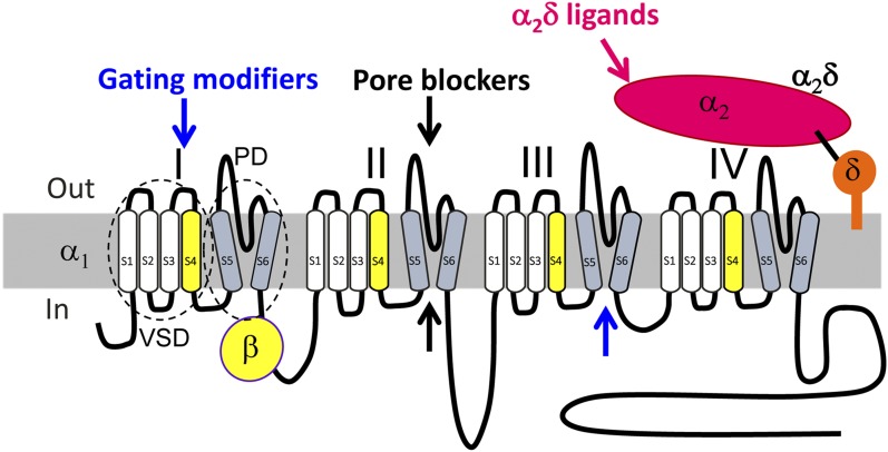
Voltage-gated calcium channels are required for many key functions in the body. In this review, the different subtypes of voltage-gated calcium channels are described and their physiologic roles and pharmacology are outlined. We describe the current uses of drugs interacting with the different calcium channel subtypes and subunits, as well as specific areas in which there is strong potential for future drug development. Current therapeutic agents include drugs targeting L-type Ca(V)1.2 calcium channels, particularly 1,4-dihydropyridines, which are widely used in the treatment of hypertension. T-type (Ca(V)3) channels are a target of ethosuximide, widely used in absence epilepsy. The auxiliary subunit α2δ-1 is the therapeutic target of the gabapentinoid drugs, which are of value in certain epilepsies and chronic neuropathic pain. The limited use of intrathecal ziconotide, a peptide blocker of N-type (Ca(V)2.2) calcium channels, as a treatment of intractable pain, gives an indication that these channels represent excellent drug targets for various pain conditions. We describe how selectivity for different subtypes of calcium channels (e.g., Ca(V)1.2 and Ca(V)1.3 L-type channels) may be achieved in the future by exploiting differences between channel isoforms in terms of sequence and biophysical properties, variation in splicing in different target tissues, and differences in the properties of the target tissues themselves in terms of membrane potential or firing frequency. Thus, use-dependent blockers of the different isoforms could selectively block calcium channels in particular pathologies, such as nociceptive neurons in pain states or in epileptic brain circuits. Of important future potential are selective Ca(V)1.3 blockers for neuropsychiatric diseases, neuroprotection in Parkinson's disease, and resistant hypertension. In addition, selective or nonselective T-type channel blockers are considered potential therapeutic targets in epilepsy, pain, obesity, sleep, and anxiety. Use-dependent N-type calcium channel blockers are likely to be of therapeutic use in chronic pain conditions. Thus, more selective calcium channel blockers hold promise for therapeutic intervention.


Figures

Fig. 1.
Fig. 1.
Diagram of voltage-gated calcium channel subunit topology. Voltage-gated calcium channel subunit topology showing major drug binding mechanisms. Channel inhibition can be induced by modification of channel gating (blue arrows, gating modifiers) by interaction with extracellular regions within one or more of the four voltage-sensing domains (VSDs) (e.g., peptide toxins, such as ω-agatoxin IVA; section III.D.2), or within the activation gates of the pore domain (PD) channel, formed by all four S5–S6 helices together (e.g., DHP LTCC blocker; section III.D). Direct block of the pore from the extracellular side (by peptide toxins such as ω-conotoxin GVIA; section II.D.2) or small molecules (with access from the cytoplasmic side) can also target regions within the ion conducting pathway and obstruct permeation through the pore (black arrows; pore blockers). Some drugs also act through both mechanisms (e.g., phenylalkylamine LTCC blockers; section II.D.1). For structural features, also see Fig. 4. The α2δ ligands (magenta arrow) can modify channel trafficking.
Fig. 2.
Fig. 2.
The most important physiologic functions of the different LTCC isoforms. Except for skeletal muscle Ca2+ channels (a complex of CaV1.1 α1 associated with β1a, α2δ-1, and γ1 subunits) and the working myocardium (CaV1.2 α1 associated with primarily β2 and α2δ-1 subunits), their subunit composition is not known for other tissues. These sites represent actual and potential sites for action of selective LTCC blockers. DA, dopamine; IHC, inner hair cells; OHCs, outer hair cells.
Fig. 3.
Fig. 3.
Modulation of L-type channels by drugs, toxins, and signaling pathways. (Pathway 1) The major pharmacologically relevant classes of LTCC active drugs are shown. DHPs (prototype nifedipine) are the most selective LTCC blockers. Verapamil and diltiazem also block non- LTCCs at higher concentrations (Diochot et al., 1995; Ishibashi et al., 1995). Tetrandrine is a bis-benzylisoquinoline alkaloid. It was isolated from the Chinese medicinal herb Stephania tetrandra used in China to treat hypertension and angina (King et al., 1988). In addition to LTCCs, it also blocks non–L-type current components (Weinsberg et al., 1994). Note that the apparent potency of classic L-type channel blockers strongly depends on membrane potential and/or stimulation frequency and action potential length. (Pathway 2) L-type channels are modulated by a variety of different signaling pathways either through membrane-delimited actions of activated G proteins (pathway 2a) or enzymes activated by GPCR (pathway 2b) or receptor tyrosine kinase (RTK) (pathway 2c) signaling (for details, see text). The FS2 structure (very similar to calciseptine) was drawn according to PDB ID 1TFS (chain trace, disulfide bonds not shown).
Fig. 4.
Fig. 4.
Calcium channel structure and ligand binding sites. (A) Extracellular view of the overall structure of CaVAb (preopen state; PDB ID 4MVQ), a homotetrameric voltage-dependent and calcium-selective channel generated by introducing three negatively charged aspartate residues (side chains in the pore are illustrated) (Tang et al., 2014). The four homologous domains of the α1 subunits of voltage-gated calcium channels likely possess a very similar architecture. Each domain contributes a voltage-sensing domain (VSD) (green, segments S1–S4) and a pore-forming domain, which together form the pore domain (PD) with a central ion conducting pathway for calcium ions (sphere). Each voltage sensor contains four positively charged arginines (side chains illustrated) that sense transmembrane voltage changes. Voltage-sensor movements are transmitted to the PD through a linker (arrow indicates one of them) between helices S4 and S5. (B) Top view of the pore module of CaVAb (pore-forming S5 and S6 helices are shown as cylinders) in the preopen state, with amino acid side chains analogous to those implicated in phenylalkylamine binding (green) and amino acid side chains specific for DHP binding illustrated in blue. The CaVAb structure has been used to illustrate how the analogs of amino acid residues important for drug binding in mammalian channels may form drug binding domains (Catterall and Swanson, 2015).The overlapping binding pocket can explain noncompetitive interactions observed in binding experiments in different tissues (Striessnig et al., 1998). (C) Top view of the CaVAb pore module in the preopen state, with the S5 and S6 segments illustrated as cylinders and amino acid side chains analogous to those implicated in phenylalkylamine binding illustrated in dark green for CaV1.2-specific residues and in light green for CaV-conserved residues. (D) Representation of DHP binding residues as in (C) for phenylalkylamines. Images in (B) to (D) were reproduced from Catterall and Swanson (2015), with permission.
Fig. 5.
Fig. 5.
Modulation of N-type channels by drugs, toxins, and signaling pathways. The major pharmacologically relevant classes of N-type calcium channel active drugs and toxins are shown in pathway 2. This includes pore-blocking peptide toxins such as ω-conotoxin MVIIA, as well as a series of different types of small organic molecules that include piperazines and piperidines, DHPs, and long-chain carbon molecules. N-type channels are modulated by a variety of different signaling pathways either through membrane-delimited actions of activated G proteins activated by GPCRs (pathway 1), or by interfering with scaffolding proteins such as CRMP-2 (pathway 3) (for details, see the text). The image of ω-conotoxin MVIIA is reproduced from Wikipedia (https://en.wikipedia.org/wiki/Ziconotide).
Fig. 6.
Fig. 6.
T-type calcium channel regulators. Examples of classes of blockers known to inhibit T-type calcium channels, including small organic molecules and the peptide toxin kurtoxin. The inhibitors either physically block the pore, or bind to the gating machinery (pathway 1). T-type calcium channels can also be regulated by activation of GPCRs, either directly by G protein βγ subunits (pathway 2a), or indirectly via protein kinases such as Rho kinase, protein kinase C, or CaMKII (pathway 2b). T-type calcium channel expression in the plasma membrane is regulated by ubiquitination and deubiquitinating. The deubiquitinase USP5 removes ubiquitin groups, thus increasing channel stability in the plasma membrane. Interfering with USP5 binding to the channel (pathway 3) leads to channel internalization and degradation. The kurtoxin image is reproduced from the Orientations of Proteins in Membranes database (Lomize et al., 2006; http://opm.phar.umich.edu/protein.php?pdbid=1t1t).

References

    1. Abbadie C, McManus OB, Sun SY, Bugianesi RM, Dai G, Haedo RJ, Herrington JB, Kaczorowski GJ, Smith MM, Swensen AM, et al. (2010) Analgesic effects of a substituted N-triazole oxindole (TROX-1), a state-dependent, voltage-gated calcium channel 2 blocker. J Pharmacol Exp Ther 334:545–555. - PubMed
    1. Adams DJ, Smith AB, Schroeder CI, Yasuda T, Lewis RJ. (2003) Omega-conotoxin CVID inhibits a pharmacologically distinct voltage-sensitive calcium channel associated with transmitter release from preganglionic nerve terminals. J Biol Chem 278:4057–4062. - PubMed
    1. Adams ME, Myers RA, Imperial JS, Olivera BM. (1993) Toxityping rat brain calcium channels with omega-toxins from spider and cone snail venoms. Biochemistry 32:12566–12570. - PubMed
    1. Adams PJ, Garcia E, David LS, Mulatz KJ, Spacey SD, Snutch TP. (2009) Ca(V)2.1 P/Q-type calcium channel alternative splicing affects the functional impact of familial hemiplegic migraine mutations: implications for calcium channelopathies. Channels (Austin) 3:110–121. - PubMed
    1. Albillos A, Neher E, Moser T. (2000) R-Type Ca2+ channels are coupled to the rapid component of secretion in mouse adrenal slice chromaffin cells. J Neurosci 20:8323–8330. - PMC - PubMed

Publication types

MeSH terms