. 2015 Sep 10;19(1):pyv083.
doi: 10.1093/ijnp/pyv083.

Hippocampal PPARδ Overexpression or Activation Represses Stress-Induced Depressive Behaviors and Enhances Neurogenesis

Affiliations

Hippocampal PPARδ Overexpression or Activation Represses Stress-Induced Depressive Behaviors and Enhances Neurogenesis

Miao-Jin Ji et al. Int J Neuropsychopharmacol. .

Erratum in

  • Erratum.
    [No authors listed] [No authors listed] Int J Neuropsychopharmacol. 2016 Apr 27;19(10):pyw031. doi: 10.1093/ijnp/pyw031. Int J Neuropsychopharmacol. 2016. PMID: 27207904 Free PMC article. No abstract available.

Abstract

Background: Emerging data have demonstrated that peroxisome proliferator-activated receptor δ (PPARδ) activation confers a potentially neuroprotective role in some neurodegenerative diseases. However, whether PPARδ is involved in depression is unknown.

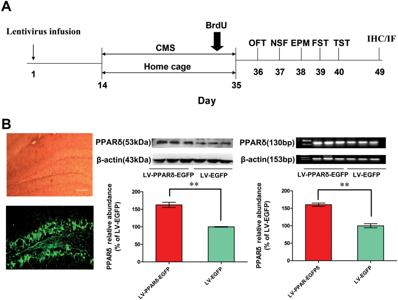
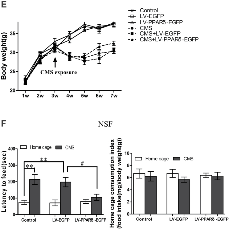
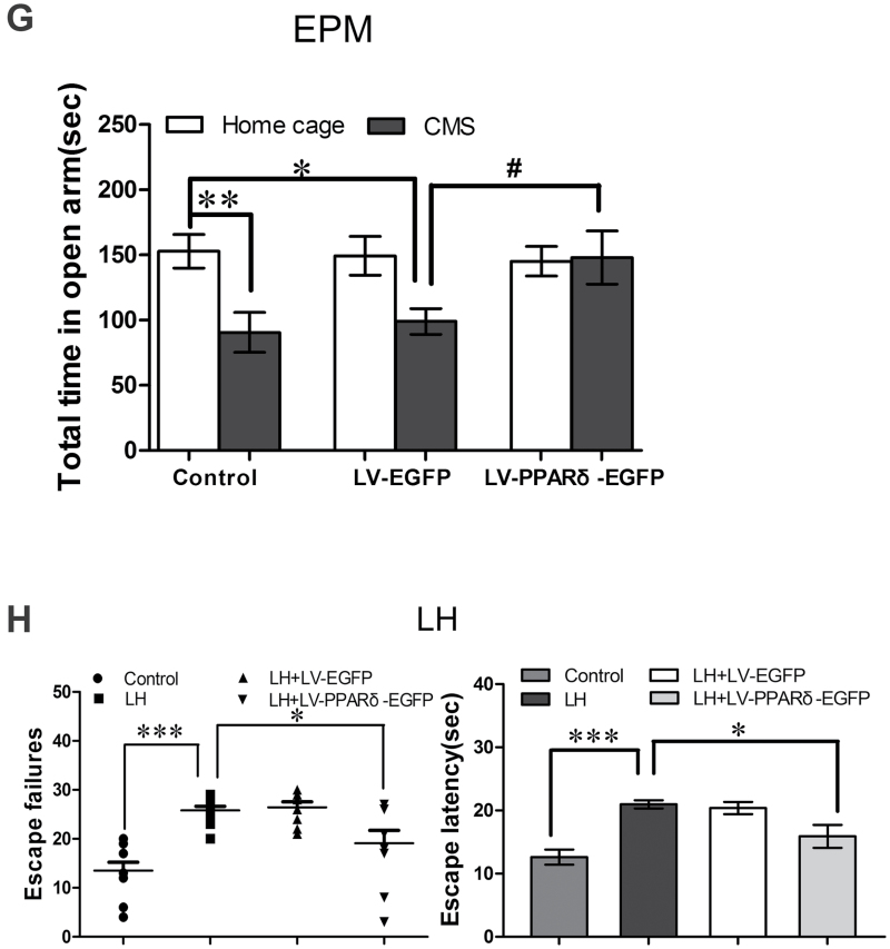
Methods: In this study, PPARδ was firstly investigated in the chronic mild stress (CMS) and learned helplessness (LH) models of depression. The changes in depressive behaviors and hippocampal neurogenesis were investigated after PPARδ overexpression by microinfusion of the lentiviral vector, containing the coding sequence of mouse PPARδ (LV-PPARδ), into the bilateral dentate gyri of the hippocampus or PPARδ activation by repeated systemic administration of PPARδ agonist GW0742 (5 or 10mg/kg.d, i.p., for 21 d).

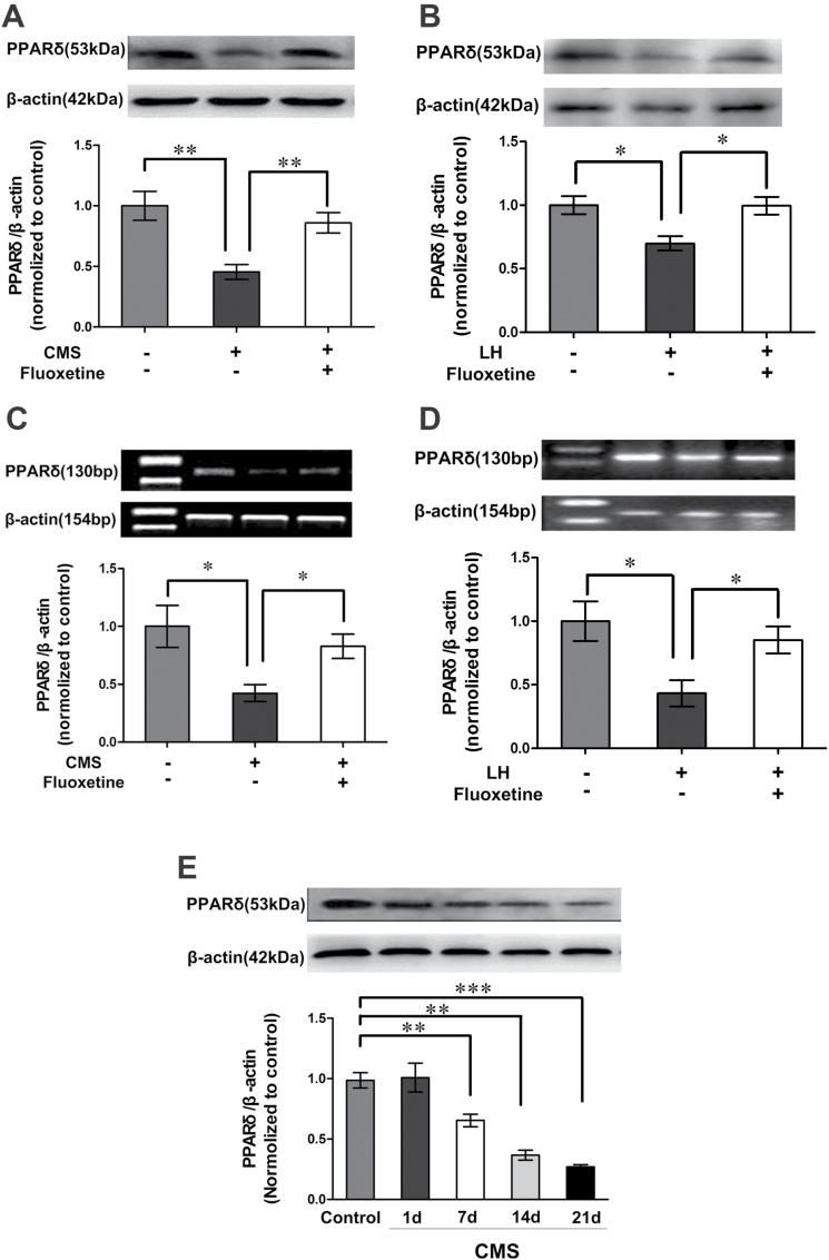
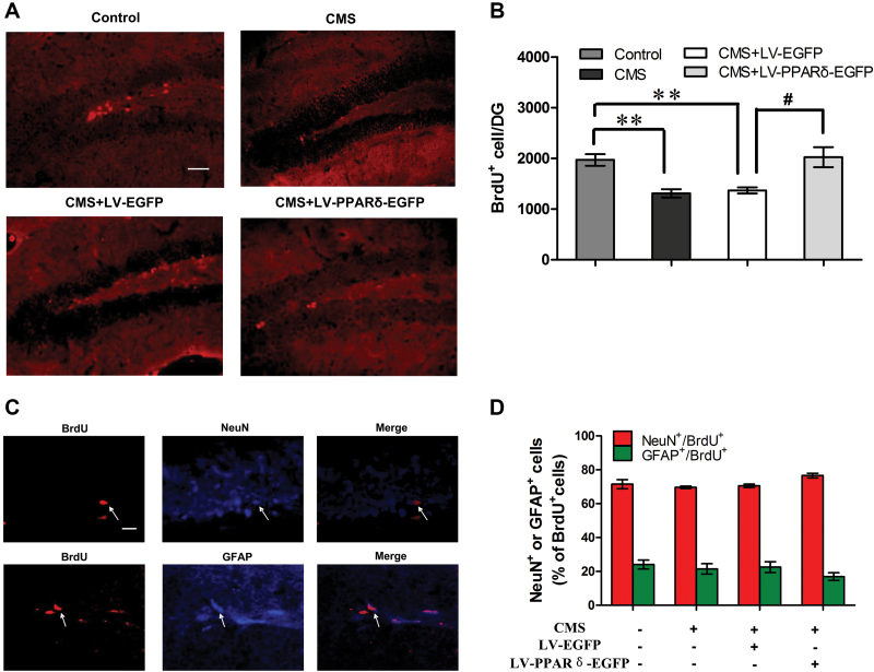
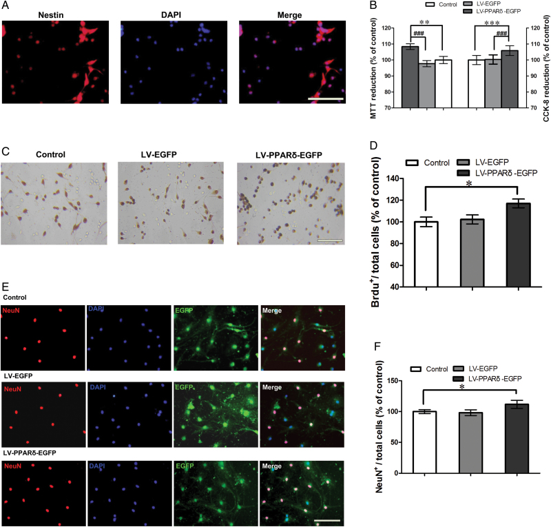
Results: We found that both CMS and LH resulted in a significant decrease in the PPARδ expression in the hippocampi of mice, and this change was reversed by treatment with the antidepressant fluoxetine. PPARδ overexpression and PPARδ activation each suppressed the CMS- and LH-induced depressive-like behavior and produced an antidepressive effect. In vivo or in vitro studies also showed that both overexpression and activation of PPARδ enhanced proliferation or differentiation of neural stem cells in the hippocampi of mice.

Conclusions: These results suggest that hippocampal PPARδ upregulation represses stress-induced depressive behaviors, accompanied by enhancement of neurogenesis.

Keywords: Depression; hippocampal neurogenesis; peroxisome proliferator-activated receptors δ.


Figures

Figure 1.
Figure 1.
Hippocampal peroxisome proliferator-activated receptor δ (PPARδ) expression is downregulated in mice exposed to chronic mild stress (CMS) or learned helplessness (LH). Immunoblots showing PPARδ protein level in the hippocampus of (A) CMS- or (B) LH-induced depression mice (n = 4). Reverse transcription-polymerase chain reaction analysis showing PPARδ mRNA in the hippocampi of (C) CMS or (D) LH depressed mice (n = 3–4). (E) Immunoblots showing hippocampal PPARδ protein of the mice treated with CMS for 1, 7, 14, or 21 d (n = 4). Data are mean ± standard error of the mean; (A, C) * p < 0.05, ** p < 0.01, compared with CMS; (B, D) * p < 0.05, ** p < 0.01, compared with LH; (E) ** p < 0.01, *** p < 0.001, compared with control.
Figure 2.
Figure 2.
Hippocampal peroxisome proliferator-activated receptor δ (PPARδ) overexpression decreases depressive behaviors. (A) Schematic timeline of the experimental procedure. OFT, open field test; NSF, novelty-suppressed feeding; EPM, elevated plus maze; FST, forced swimming test; TST, tail suspension test; IHC, immunohistochemistry; IF, immunofluorescence. (B) Shown are representative dentate gyrus (DG) area with transfection of a lentiviral vector that selectively expresses PPARδ with enhanced green fluorescent protein (LV-PPARδ-EGFP), PPARδ protein, or mRNA in the hippocampus (n = 3). Shown are the (C) immobility time in the forced swim test (FST) and tail suspension test (TST), (D) physical state index, (E) body weight, (F) latency to feed and home cage consumption index in NSF test, and (G) total time spent in open arms in EPM in the mice intrahippocampally microinjected with LV-PPARδ-EGFP (2×10⁹ TU, 2μl/side), followed 2 weeks later by chronic mild stress (CMS) for 21 d (n = 8–10). (H) Shown are escape failures and escape latency in the learned helplessness (LH) test (n = 8–10). Data are mean±standard error of the mean. (B) ** p < 0.01 compared with a lentiviral vector expressing EGFP alone (LV-EGFP); (C-G)* p < 0.05, ** p < 0.01 compared with control; # p < 0.05, ## p < 0.01, compared with CMS plus LV-EGFP; (H) * p < 0.05, ** p < 0.01 compared with LH.
Figure 2.
Figure 2.
Hippocampal peroxisome proliferator-activated receptor δ (PPARδ) overexpression decreases depressive behaviors. (A) Schematic timeline of the experimental procedure. OFT, open field test; NSF, novelty-suppressed feeding; EPM, elevated plus maze; FST, forced swimming test; TST, tail suspension test; IHC, immunohistochemistry; IF, immunofluorescence. (B) Shown are representative dentate gyrus (DG) area with transfection of a lentiviral vector that selectively expresses PPARδ with enhanced green fluorescent protein (LV-PPARδ-EGFP), PPARδ protein, or mRNA in the hippocampus (n = 3). Shown are the (C) immobility time in the forced swim test (FST) and tail suspension test (TST), (D) physical state index, (E) body weight, (F) latency to feed and home cage consumption index in NSF test, and (G) total time spent in open arms in EPM in the mice intrahippocampally microinjected with LV-PPARδ-EGFP (2×10⁹ TU, 2μl/side), followed 2 weeks later by chronic mild stress (CMS) for 21 d (n = 8–10). (H) Shown are escape failures and escape latency in the learned helplessness (LH) test (n = 8–10). Data are mean±standard error of the mean. (B) ** p < 0.01 compared with a lentiviral vector expressing EGFP alone (LV-EGFP); (C-G)* p < 0.05, ** p < 0.01 compared with control; # p < 0.05, ## p < 0.01, compared with CMS plus LV-EGFP; (H) * p < 0.05, ** p < 0.01 compared with LH.
Figure 3.
Figure 3.
Peroxisome proliferator-activated receptor δ (PPARδ) overexpression enhances hippocampal neurogenesis. (A) Representative cells and(B) quantification of bromodeoxyuridine (BrdU)-labeled cells (red) in the dentate gyrus (DG) from control or chronic mild stress (CMS) mice with a lentiviral vector expressing enhanced green fluorescent protein alone (LV-EGFP) or a lentiviral vector that selectively expresses PPARδ with EGFP (LV-PPARδ-EGFP; n = 4). Scale bars = 50 μm. (C) Phenotype of BrdU-positive cells in the DG of CMS + LV-PPARδ-EGFP mice, and higher magnification of cells double-labeled for BrdU (red; left) and the neuronal marker nuclear antigen (NeuN; blue, middle and top) or the glialmarker glial fibrillary acidic protein (GFAP; blue, middle and bottom). Scale bars = 20 μm. (D) Percentages of neuronal and glial cells labeled by BrdU in the DG of control or CMS mice treated with LV-EGFP or LV-PPARδ-EGFP (n = 4). Data are mean ± standard error of the mean. * p < 0.01, compared with control, # p < 0.05, compared with CMS plus LV-EGFP.
Figure 4.
Figure 4.
Peroxisome proliferator-activated receptor δ (PPARδ) overexpression promoted proliferation and differentiation of neural stem cells (NSCs) in vitro. (A) Shown is the protein marker nestin, expressed by NSCs. (B) The coding sequence of mouse PPARδ (LV-PPARδ) increases proliferation determined by 3-(4, 5-dimethythiazole-2-yl)-2, 5-diphenyl-tetrazolium bromide (MTT) and cell counting kit (CCK-8) assays (n = 6). (C) Representatives of bromodeoxyuridine positive (BrdU+)-labeled cells of the NSCs after treatment with a lentiviral vector that selectively expresses PPARδ with enhanced green fluorescent protein (LV-PPARδ-EGFP). (D) Statistical graph shows the number of BrdU+ cells in different groups (n = 6). (E) Immunocytochemistry for neuronal marker neuronal nuclear antigen (NeuN) was used to assess neural differentiation in NSCs treated with LV-PPARδ. (F) Shown are statistical data of NeuN+-positive neurons in different groups (n = 6). Data are mean ± standard error of the mean. * p < 0.05, ** p < 0.01, *** p < 0.001, compared with control; # p < 0.001, compared with a lentiviral vector expressing EGFP alone (LV-EGFP). Scale bars = 50 μm.

References

    1. Arsenijevic D, de Bilbao F, Plamondon J, Paradis E, Vallet P, Richard D, Lanqhans W, Giannakopoulos P. (2006) Increased infarct size and lack of hyperphagic response after focal cerebral ischemia in peroxisome proliferator-activated receptor beta-deficient mice. J Cereb Blood Flow Metab 26:433–445. - PubMed
    1. Bartolomucci A, Leopardi R. (2009) Stress and depression: preclinical research and clinical implications. PLOS ONE 4:e4265. - PMC - PubMed
    1. Belmaker RH, Agam G. (2008) Major depressive disorder. N Engl J Med 358:55–68. - PubMed
    1. Braissant O, Foufelle F, Scotto C, Dauça M, Wahli W. (1996) Differential expression of peroxisome proliferator-activated receptors (PPARs): tissue distribution of PPAR-alpha, -beta, and -gamma in the adult rat. Endocrinology 137:354–366. - PubMed
    1. Caldarone BJ, George TP, Zachariou V, Picciotto MR. (2000) Gender differences in learned helplessness behavior are influenced by genetic background. Pharmacol Biochem Behav 66:811–817. - PubMed

Publication types

MeSH terms

Substances