. 2015 Oct 15;195(8):4010-9.
doi: 10.4049/jimmunol.1500447. Epub 2015 Sep 11.

NK Cells Preferentially Target Tumor Cells with a Cancer Stem Cell Phenotype

Affiliations

NK Cells Preferentially Target Tumor Cells with a Cancer Stem Cell Phenotype

Erik Ames et al. J Immunol. .

Abstract

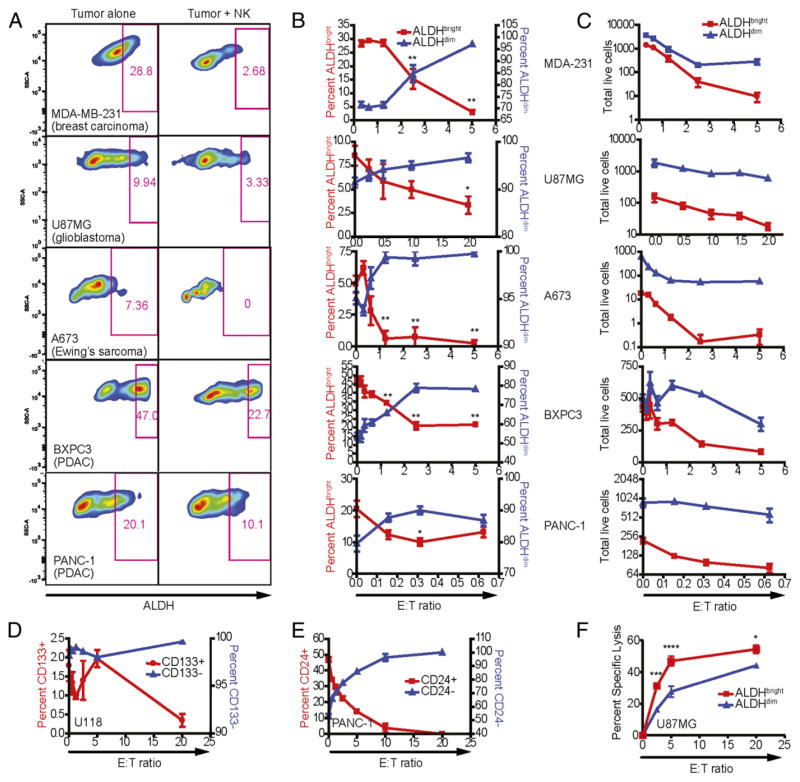
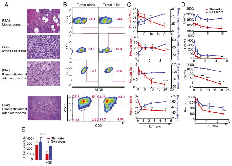
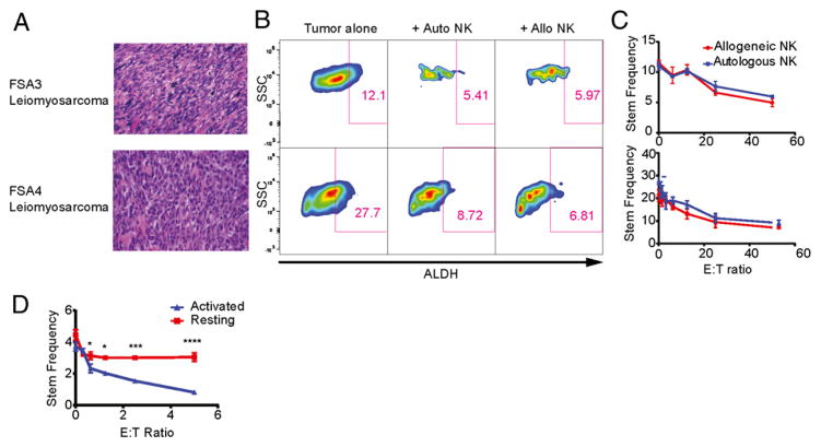
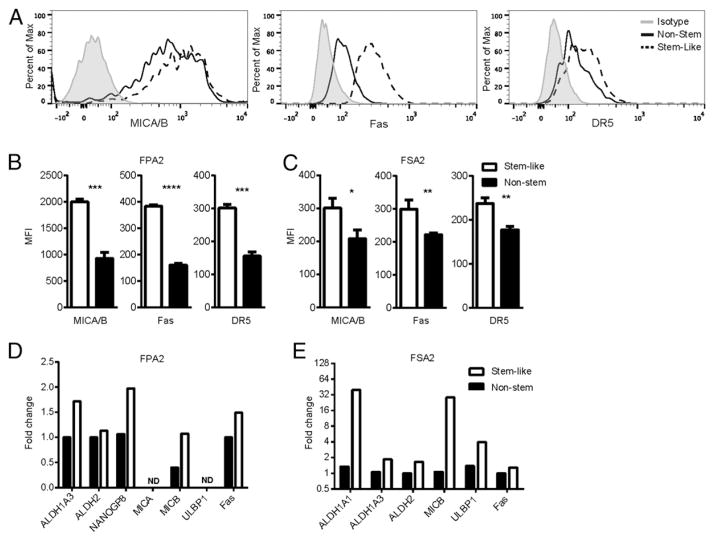
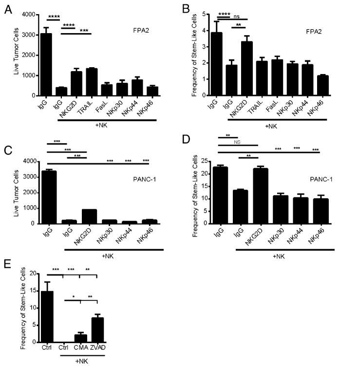
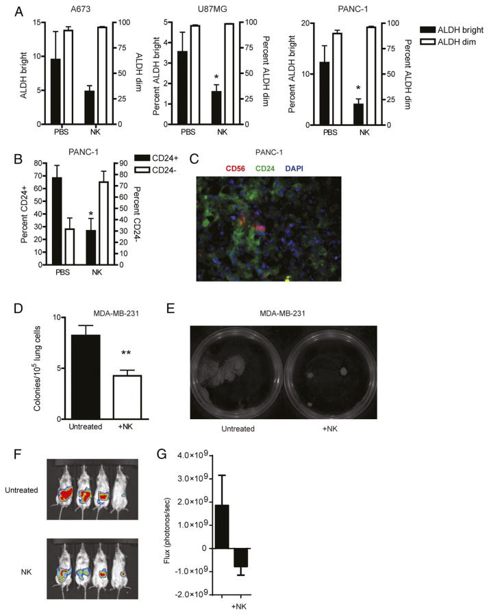
Increasing evidence supports the hypothesis that cancer stem cells (CSCs) are resistant to antiproliferative therapies, able to repopulate tumor bulk, and seed metastasis. NK cells are able to target stem cells as shown by their ability to reject allogeneic hematopoietic stem cells but not solid tissue grafts. Using multiple preclinical models, including NK coculture (autologous and allogeneic) with multiple human cancer cell lines and dissociated primary cancer specimens and NK transfer in NSG mice harboring orthotopic pancreatic cancer xenografts, we assessed CSC viability, CSC frequency, expression of death receptor ligands, and tumor burden. We demonstrate that activated NK cells are capable of preferentially killing CSCs identified by multiple CSC markers (CD24(+)/CD44(+), CD133(+), and aldehyde dehydrogenase(bright)) from a wide variety of human cancer cell lines in vitro and dissociated primary cancer specimens ex vivo. We observed comparable effector function of allogeneic and autologous NK cells. We also observed preferential upregulation of NK activation ligands MICA/B, Fas, and DR5 on CSCs. Blocking studies further implicated an NKG2D-dependent mechanism for NK killing of CSCs. Treatment of orthotopic human pancreatic cancer tumor-bearing NSG mice with activated NK cells led to significant reductions in both intratumoral CSCs and tumor burden. Taken together, these data from multiple preclinical models, including a strong reliance on primary human cancer specimens, provide compelling preclinical evidence that activated NK cells preferentially target cancer cells with a CSC phenotype, highlighting the translational potential of NK immunotherapy as part of a combined modality approach for refractory solid malignancies.


Conflict of interest statement

Disclosures

The authors have no financial conflicts of interest.

Figures

FIGURE 1
FIGURE 1
Sensitivity of human tumor cell lines to NK cell lysis. Human NK cells were activated with IL-2, expanded for 2 wk, and cocultured 16–18 h with MDA-MB-231 (breast adenocarcinoma), U87MG (glioblastoma), A673 (Ewing’s sarcoma), BxPC3 (pancreatic ductal adenocarcinoma), and PANC-1 (pancreatic ductal adenocarcinoma) in the presence of 500 IU/ml rhIL-2. Phenotypes of remaining tumor cells were assessed by excluding 7-AAD+ events. (A) ALDH expression was assessed using the Aldefluor system in cultures with tumor cells alone and with NK cells at E:T ratios between 1:1 and 5:1. (B) The percentage of ALDHbright events on tumor cells was plotted (red; left y-axis) against the ALDHdim population (blue; right y-axis) at the indicated E:T ratios. (C) The total number of acquired events from (B) were plotted over the indicated E:T ratios. Reductions in ALDHbright populations in comparison with untreated cells were determined via one-way ANOVA with a Tukey posttest. *p < 0.05; **p < 0.01. (D) CSCs from the U118 glioblastoma were identified based on expression of surface marker CD133 (glioblastoma) following culture with NK cells at the specified E:T ratios. (E) Changes in the proportions of CD24-expressing and nonexpressing PANC-1 cells following NK coculture. (F) U87-MG cells were sorted for ALDH expression and then assessed for sensitivity to NK cell cytotoxicity in a 4-h 51Cr-release assay.
FIGURE 2
FIGURE 2
CSCs from human primary tumors are preferentially targeted by human NK cells. (A) Well-differentiated liposarcoma (FSA1), Ewing’s sarcoma (FSA2), and pancreatic ductal adenocarcinoma (FPA1 and FPA2) were obtained from fresh clinical specimens (H&E staining, original magnification ×40). (B) ALDH or CD24/CD44 expression on tumor targets was assessed with tumor cells alone or those cocultured with NK cells at E:T ratios between 1:1 and 5:1. (C) The percentage of CSC populations on tumor cells was plotted (red; left y-axis) against the non-CSC population (blue; right y-axis) at the indicated E:T ratios. (D) The total number of acquired events from (B) were plotted over the indicated E:T ratios. (E) Stem-like (CD24+/CD44+/ALDHbright) or non–stem cells (CD24/ALDHdim) from FPA2 were sorted then used in a 16-h NK cell cytotoxicity assay with allogeneic NK cells. Reductions in CSC populations were determined via one-way ANOVA with a Tukey posttest. *p < 0.05, ** p < 0.01, ***p < 0.001, ****p < 0.0001.
FIGURE 3
FIGURE 3
Autologous NK cells can effectively kill primary CSCs. (A) Fresh primary leiomyosarcomas FSA3 and FSA4 were processed into single-cell suspensions and used in cytotoxicity assays with either patient-matched autologous or healthy donor allogeneic NK cells (H&E staining, original magnification ×40). (B) ALDH expression of tumor cells was assessed by excluding CD45+ and 7-AAD+ events. (C) The percentages of ALDHbright tumor cells were determined following coculture with either autologous or allogeneic NK cells at E:T ratios ranging from 0 to 50. (D) NK cells were isolated from a patient both on the day of the resection of a leiomyosarcoma or 7 d before. NK cells isolated 7 d before the resection were activated with IL-2 as before whereas those isolated on the same day as the tumor were left unactivated. Both batches of NK cells were cultured with tumor cell suspensions in a flow cytometric cytotoxicity assay at the indicated E:T ratios. The percentage of remaining tumor cells showing a CSC-like phenotype via ALDH expression are shown and differences between resting and activated NK cells were determined via two-way ANOVA with a Sidak multiple comparisons test. *p < 0.05, ***p < 0.001, ****p < 0.0001.
FIGURE 4
FIGURE 4
CSCs express higher levels of NKG2D ligands and death receptors than do non-CSCs. (A) Histograms of median fluorescence intensity (MFI) of MICA/B, Fas, and DR5 by flow cytometry are shown from CSC (dotted lines) and non-CSC (solid lines) populations from the primary pancreatic adenocarcinoma, FPA2. Quantified MFI values are shown for FPA2 (B) and a soft-tissue sarcoma (FSA2) (C). CSC populations from these tumor specimens were sorted and assessed by quantitative RT-PCR for CSC markers and stress ligands on FPA2 (D) and FSA2 (E). *p < 0.05, **p < 0.01, ***p < 0.001.
FIGURE 5
FIGURE 5
Blocking NKG2D abrogates NK killing of CSCs from cell lines and primary tumors. FPA2 (A and B) or PANC-1 (C and D) cells were cultured with activated NK cells (1:1 E:T ratio) for 16 h in the presence of the indicated Fc-chimeric proteins that blocked the indicated activation receptors. Human IgG1 purified Ab was added as an isotype-matched control. Tumors were then analyzed by flow cytometry for total numbers of remaining live tumor cells (SSChiCD457-AAD) (A and C) and percentages of tumor cells expressing CD24/CD44/ALDHbright (B and D). (E) PANC-1 cells were pretreated with 80 μM ZVAD or vehicle control for 1 h, or NK cells were pretreated with 50 nM CMA for 1 h. NK cells and PANC-1 tumor cells were then mixed at a 5:1 ratio and ZVAD, CMA, or vehicle controls were adjusted to maintain the above-stated concentrations. Twenty-four hours later, wells were harvested and analyzed for ALDH expression by flow cytometry. Reductions in stem-like populations in comparison with untreated cells were determined via one-way ANOVA with a Tukey posttest. *p < 0.05, **p < 0.01, ***p < 0.001, ****p < 0.0001.
FIGURE 6
FIGURE 6
NK cells attack CSCs in vivo. Mice bearing s.c. A673, U87, and PANC-1 tumors were intratumorally injected with 2 × 10⁷ activated NK cells. Six days later, mice were sacrificed and tumors were processed into single-cell suspensions and assessed for ALDH (A) or CD24 (B) expression. ALDHbright or CD24 (CSC) percentages are plotted on the left y-axis and ALDHdim or CD24 (non-CSC) percentages are plotted on the right y-axis. (C) Colocalization of transferred NK cells and CD24-expressing CSCs was detected using immunofluorescence (anti-CD56, red; anti-CD24, green; DAPI, blue; original magnification ×40). (D) Lungs from mice bearing metastatic MDA-MB-231 tumors were plated in CFU assays 5 d after in vivo NK cell injections. The number of tumor colonies were counted (D) and imaged for comparison (E). NSG mice were given orthotopic injections of PANC-1 into the pancreas. Twenty days following tumor implantation, 2 × 10⁷ activated NK cells were injected i.v. Bioluminescent images (F) and combined values (G) were assessed 14 d after NK cell transfer. Experiments were performed twice with three to four mice per group. Statistics were determined by one-way ANOVA with a Bonferroni posttest (A and B) or Student t test (C, D, and G). *p < 0.05, **p < 0.01.

References

    1. Visvader JE, Lindeman GJ. Cancer stem cells: current status and evolving complexities. Cell Stem Cell. 2012;10:717–728. - PubMed
    1. Chen J, Li Y, Yu TS, McKay RM, Burns DK, Kernie SG, Parada LF. A restricted cell population propagates glioblastoma growth after chemotherapy. Nature. 2012;488:522–526. - PMC - PubMed
    1. Valent P, Bonnet D, De Maria R, Lapidot T, Copland M, Melo JV, Chomienne C, Ishikawa F, Schuringa JJ, Stassi G, et al. Cancer stem cell definitions and terminology: the devil is in the details. Nat Rev Cancer. 2012;12:767–775. - PubMed
    1. Driessens G, Beck B, Caauwe A, Simons BD, Blanpain C. Defining the mode of tumour growth by clonal analysis. Nature. 2012;488:527–530. - PMC - PubMed
    1. Wang L, Park P, Zhang H, La Marca F, Lin CY. Prospective identification of tumorigenic osteosarcoma cancer stem cells in OS99-1 cells based on high aldehyde dehydrogenase activity. Int J Cancer. 2011;128:294–303. - PubMed

Publication types