. 2015 Nov 6;290(45):26943-26953.
doi: 10.1074/jbc.M115.682138. Epub 2015 Sep 11.

Novel Insights into Interleukin 6 (IL-6) Cis- and Trans-signaling Pathways by Differentially Manipulating the Assembly of the IL-6 Signaling Complex

Affiliations

Novel Insights into Interleukin 6 (IL-6) Cis- and Trans-signaling Pathways by Differentially Manipulating the Assembly of the IL-6 Signaling Complex

Marine Lacroix et al. J Biol Chem. .

Abstract

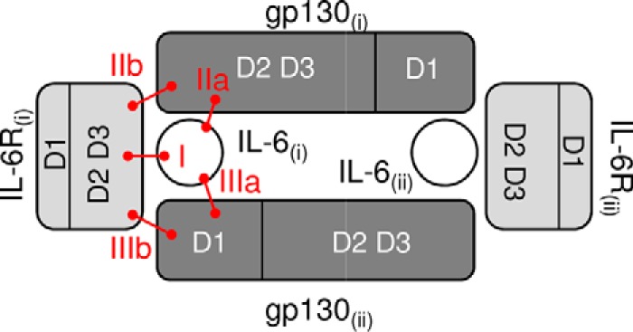
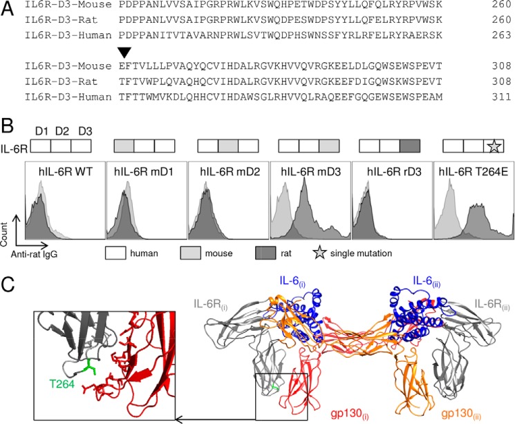
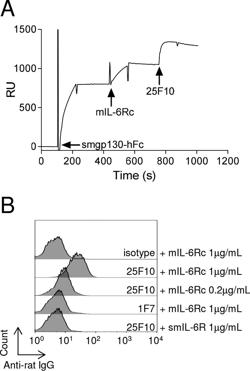
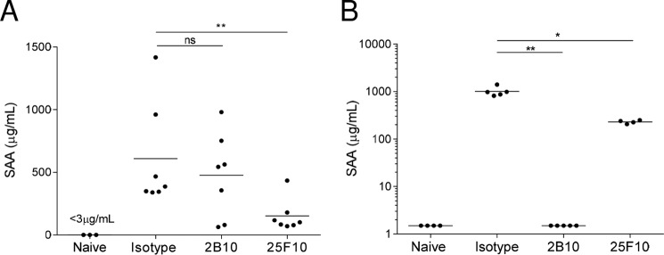
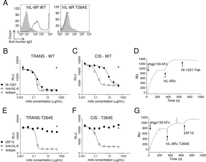
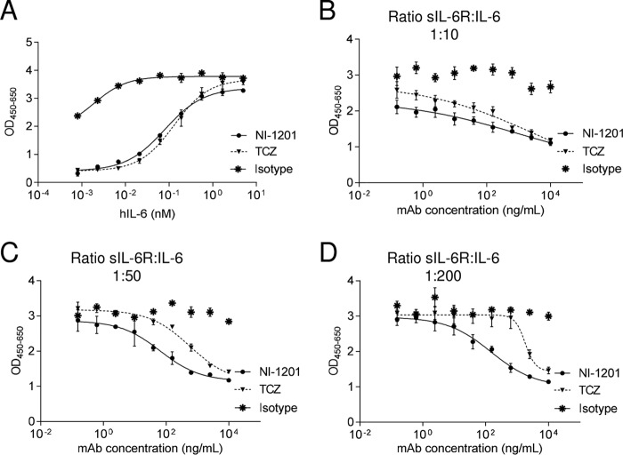
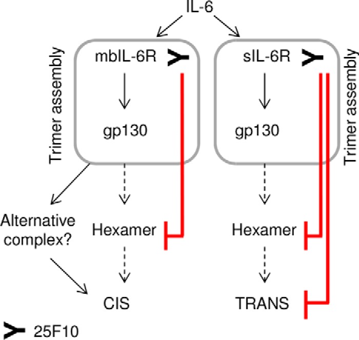
The IL-6 signaling complex is described as a hexamer, formed by the association of two IL-6·IL-6 receptor (IL-6R)·gp130 trimers, with gp130 being the signal transducer inducing cis- and trans-mediated signaling via a membrane-bound or soluble form of the IL-6R, respectively. 25F10 is an anti-mouse IL-6R mAb that binds to both membrane-bound IL-6R and soluble IL-6R with the unique property of specifically inhibiting trans-mediated signaling events. In this study, epitope mapping revealed that 25F10 interacts at site IIb of IL-6R but allows the binding of IL-6 to the IL-6R and the recruitment of gp130, forming a trimer complex. Binding of 25F10 to IL-6R prevented the formation of the hexameric complex obligate for trans-mediated signaling, suggesting that the cis- and trans-modes of IL-6 signaling adopt different mechanisms for receptor complex assembly. To study this phenomenon also in the human system, we developed NI-1201, a mAb that targets, in the human IL-6R sequence, the epitope recognized by 25F10 for mice. Interestingly, NI-1201, however, did not selectively inhibit human IL-6 trans-signaling, although both mAbs produced beneficial outcomes in conditions of exacerbated IL-6 as compared with a site I-directed mAb. These findings shed light on the complexity of IL-6 signaling. First, triggering cis- versus trans-mediated IL-6 signaling occurs via distinctive mechanisms for receptor complex assembly in mice. Second, the formation of the receptor complex leading to cis- and trans-signaling biology in mice and humans is different, and this should be taken into account when developing strategies to inhibit IL-6 clinically.

Keywords: antibody; complex; interleukin 6 (IL-6); receptor; signaling.


Figures

FIGURE 1.
FIGURE 1.
Schematic view of the interacting domains within the IL-6 hexameric signaling complex. IL-6 interacts with D2 and D3 of IL-6R (site I). Within this dimer, IL-6 and IL-6R are both involved in binding to D2 and D3 of gp130 through sites IIa and IIb, respectively. Additional interactions form the IL-6 signaling hexameric complex by assembling two dimers (i and ii) of IL-6·IL-6R·gp130 through D1 of gp130 (sites IIIa and IIIb). IL-6 is in white, IL-6R is in light gray, and gp130 is in dark gray.
FIGURE 2.
FIGURE 2.
25F10 engages mbIL-6R but does not inhibit IL-6 cis-signaling, only trans-signaling. A, STAT3-luciferase-transfected PEAK cells were stimulated with mIL-6 (0.5 μg/ml) and smIL-6R (1 μg/ml), and after 18 h, firefly activity was measured in the presence or absence of varying concentrations of 25F10 or 2B10. mIL-6 and smIL-6R alone were used as negative controls. mIL-6 in combination with smIL-6R and an isotype control mAb at 0.67 × 10⁻⁸ m was used as a positive control. Error bars represent S.E. B, IL-6R+ T1165 cells were incubated for 30 min with 25F10, 2B10, or isotype control. Binding was assessed by flow cytometry. C, T1165 cells were stimulated with IL-6 (1 ng/ml), and after 48 h, cell proliferation was measured in the presence or absence of varying concentrations of 25F10 or 2B10. The results are presented as a percentage of proliferation normalized to the signal induced by IL-6 only. Error bars represent S.E. RLU, relative light units.
FIGURE 3.
FIGURE 3.
25F10 binds the interaction site IIb of the IL-6 signaling complex. A, sequence alignment of D3 of mouse, rat, and human IL-6Rs. The residue responsible for the binding of 25F10 is indicated (▾). B, PEAK cells were transiently transfected with chimeric or mutated hIL-6R as indicated in each panel. 25F10 or isotype control was added to the cells, and mAbs bound to the surface were detected with an APC-coupled anti-rat IgG and analyzed by flow cytometry (25F10, filled dark gray; isotype control, filled light gray). Results are representative of at least two independent experiments. C, schematic view of the human IL-6 signaling hexameric complex generated with PyMOL software (Protein Data Bank code 1P9M) highlighting in green the amino acid responsible for the binding of 25F10 among its murine equivalent within the site IIb. The enlargement is a view of site IIb shown at higher magnification depicting Thr-264 among IL-6R and residues of gp130 involved in this interface (17).
FIGURE 4.
FIGURE 4.
The trimer IL-6·IL-6R·gp130 assembles even in the presence of 25F10. A, an anti-human Fc was immobilized on a CM5 chip. Smgp130-hFc, mIL-6Rc, and 25F10 were sequentially injected at 10, 5, and 100 μg/ml, respectively. The sensorgram signal is shown as relative units (RU). Arrows indicate the start of an injection. Results are representative of at least two independent experiments. B, mIL-6Rc or smIL-6R (both at 1 μg/ml) was premixed with 5 μg/ml 25F10, isotype control, or 1F7, a rat anti-mIL-6R mAb that blocks the interaction between IL-6·IL-6R and gp130, prior to incubation for 15 min at 4 °C with NIH3T3 cells. mAbs bound to the cell surface were detected using an APC-coupled anti-rat IgG antibody, and cells were analyzed by flow cytometry.
FIGURE 5.
FIGURE 5.
In the presence of 25F10, a tetrameric complex, i.e. IL-6·IL-6R·25F10·gp130, is formed in vivo. A, schematic view of the different ways of target-driven elimination of 25F10 in WT versus IL-6−/− mice. In WT mice, 25F10 interacts with its targets mbIL-6R and sIL-6R and allows the formation of four distinct complexes at the cell membrane that drive the elimination of 25F10 from the circulation. In IL-6−/− mice, target-driven elimination of 25F10 is only able to be mediated by mbIL-6R as the other formats shown in the top panel cannot form in the absence of IL-6. B, 25F10 mAb was intravenously administered to WT or IL-6−/− mice at 10 mg/kg. Plasma samples were obtained at the indicated time points following injection, starting at 1 h postdosing, and the mAb concentration was analyzed by ELISA. Data are expressed as the mean ± S.E. (error bars) (n = 3–4 mice). mb, cell membrane; LLOD, lower limit of detection of the assay.
FIGURE 6.
FIGURE 6.
In vivo IL-6 signaling blockade with 25F10 affords enhanced abrogation of IL-6 responses under inflammatory conditions. A, mice were injected with 50 μg of CpG-ODN on days 0, 2, 4, and 7. Mice were treated with mAbs intravenously at 10 mg/kg simultaneously with the last CpG injection (on day 7). Plasma was harvested 24 h after the last CpG injection, and SAA plasma concentrations were measured by ELISA. B, mice were treated with mAbs intravenously at 10 mg/kg simultaneously with the CFA intradermic injection. Plasma was harvested 24 h after the injections, and SAA plasma concentrations were measured by ELISA. Data are expressed as the mean. Statistical analyses were performed between the indicated group and the isotype control group values. ns, not significant; *, p < 0.05; **, p < 0.01 were obtained using the one-tailed non-parametric Mann-Whitney U test.
FIGURE 7.
FIGURE 7.
NI-1201 targets the same epitope on the human protein as 25F10 does for the mouse receptor but inhibits both IL-6 cis- and trans-signaling. A, PEAK cells were transiently transfected with wild-type or mutated hIL-6R as indicated in each panel. NI-1201 or isotype control was added to the cells, and mAbs bound to the surface were detected with anti-human IgG-APC and analyzed by flow cytometry (NI-1201, black line; isotype control, filled gray). Results are representative of at least two independent experiments. B and E, PEAK cells were transfected with pSIEM-STAT3-luciferase vector and incubated with serial dilutions of mAbs or isotype control as indicated and with 100 ng/ml hIL-6Rc WT or hIL-6Rc-T264E. Firefly luciferase activity was monitored 16 h later. C and F, PEAK cells were transfected with a vector containing hIL-6R or hIL-6R-T264E and 1 day later with pSIEM-STAT3-luciferase vector. They were then co-incubated with serial dilutions of mAbs or isotype control as indicated and with 10 ng/ml hIL-6. Data are expressed as the mean ± S.E. (error bars) and are representative of four independent experiments. D, anti-human Fc was immobilized on a CM5 chip. Shgp130-hFc, hIL-6Rc WT, and NI-1201 Fab were sequentially injected at 10, 10, and 50 μg/ml, respectively. Arrows indicate the start of the injection. Results are representative of at least two independent experiments. G, anti-human Fc was immobilized on a CM5 chip. Shgp130-hFc, mutated hIL-6Rc-T264E, and 25F10 were sequentially injected at 10, 10, and 5 μg/ml, respectively. Arrows indicate the start of an injection. Results are representative of at least two independent experiments. RLU, relative light units; RU, relative units.
FIGURE 8.
FIGURE 8.
NI-1201 abrogates more efficiently IL-6 trans-signaling activity in conditions of exacerbated IL-6 compared with tocilizumab. A, Ba/F3-hgp130-hIL-6R cells were stimulated with increasing concentrations of hIL-6 and 10 nm NI-1201 or tocilizumab. After 72 h, cell proliferation was measured. Data are expressed as the mean ± S.E. B–D, Ba/F3-hgp130 cells were stimulated with increasing hIL-6 concentrations and a fixed shIL-6R concentration (10 ng/ml) at the indicated molar ratios. After 72 h, cell proliferation was measured in the presence or absence of varying concentrations of NI-1201, tocilizumab, or isotype control. Data are expressed as the mean ± S.E. (error bars). TCZ, tocilizumab.
FIGURE 9.
FIGURE 9.
Model to illustrate the potential mechanism of action of 25F10. 25F10 inhibits the hexamer assembly when bound to mb- and sIL-6R (red lines). Nevertheless, the IL-6·mbIL-6R·25F10·gp130 complex may be considered as an intermediary step to induce cis-signaling, whereas hexamer formation would be sufficient and necessary for trans-signaling.

References

    1. Mihara M., Hashizume M., Yoshida H., Suzuki M., and Shiina M. (2012) IL-6/IL-6 receptor system and its role in physiological and pathological conditions. Clin. Sci. 122, 143–159 - PubMed
    1. Peters M., Müller A. M., and Rose-John S. (1998) Interleukin-6 and soluble interleukin-6 receptor: direct stimulation of gp130 and hematopoiesis. Blood 92, 3495–3504 - PubMed
    1. Scheller J., Chalaris A., Schmidt-Arras D., and Rose-John S. (2011) The pro- and anti-inflammatory properties of the cytokine interleukin-6. Biochim. Biophys. Acta 1813, 878–888 - PubMed
    1. Scheller J., Garbers C., and Rose-John S. (2014) Interleukin-6: from basic biology to selective blockade of pro-inflammatory activities. Semin. Immunol. 26, 2–12 - PubMed
    1. Heinrich P. C., Behrmann I., Haan S., Hermanns H. M., Müller-Newen G., and Schaper F. (2003) Principles of interleukin (IL)-6-type cytokine signalling and its regulation. Biochem. J. 374, 1–20 - PMC - PubMed

MeSH terms

Associated data