Review
. 2015 Oct;24(4):795-832.
doi: 10.1016/j.soc.2015.06.008. Epub 2015 Jul 27.

Multiple Endocrine Neoplasia: Genetics and Clinical Management

Affiliations
Review

Multiple Endocrine Neoplasia: Genetics and Clinical Management

Jeffrey A Norton et al. Surg Oncol Clin N Am. 2015 Oct.

Abstract

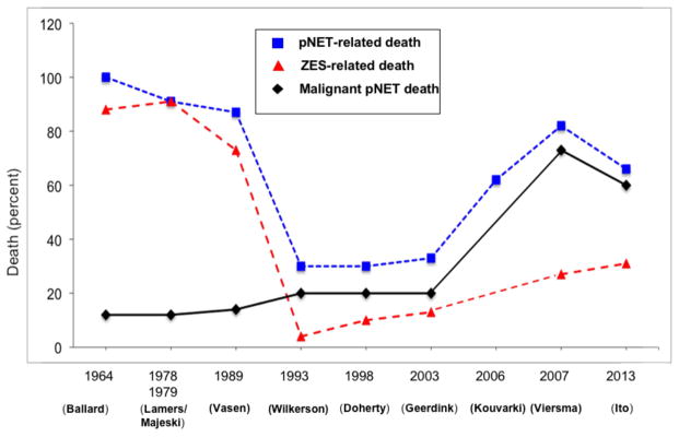
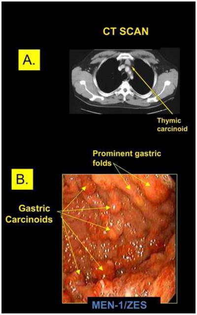
Early diagnosis of multiple endocrine neoplasia (MEN) syndromes is critical for optimal clinical outcomes; before the MEN syndromes can be diagnosed, they must be suspected. Genetic testing for germline alterations in both the MEN type 1 (MEN1) gene and RET proto-oncogene is crucial to identifying those at risk in affected kindreds and directing timely surveillance and surgical therapy to those at greatest risk of potentially life-threatening neoplasia. Pancreatic, thymic, and bronchial neuroendocrine tumors are the leading cause of death in patients with MEN1 and should be aggressively considered by at least biannual computed tomography imaging.

Keywords: Clinical management; Endocrine neoplasia; Genetics; Multiple endocrine neoplasia.


Figures

Figure 1
Figure 1. Sestamibi scan (A), neck CT scan (B) and somatostatin receptor scintigraphy SRS (C) in an MEN-1 patient with recurrent primary hyperparathryoidism and metastatic pancreatic NET
Patient is a 45 year old man with recurrent primary hyperparathyroidism status post an operation in which they removed two abnormal parathyroid glands and now he has recurrent disease. Sestamibi scan (A) shows an abnormal right inferior parathyroid gland (yellow arrow) and the same abnormal gland is demonstrated on a 4-D neck CT scan with intravenous contrast (yellow arrow) (B). Octreoscan shows an abnormal pancreatic NET in the tail of the pancreas with possible liver metastases (C) left at 4 hours and middle panel 24 hours after labelled octreotide), while 68Ga DOTATATE (C right panel) clearly shows more tumor and also a pituitary adenoma.
Figure 2
Figure 2. Histologic appearance of the pancreas in two separate patients with MEN-1
(A) Histologic sections of a pancreas in a patient with multiple neuroendocrine microadenomas in a background of islet cell hyperplasia. Panel 1 (40X): The H&E stain demonstrates clusters of enlarged microadenomas, the largest of which is 0.35 cm. Panel 2 (40X): Immunhistochemical staining for synaptophysin highlights the multiple (>4) neuroendocrine microadenomas. (B) Histologic sections of a pancreatic neuroendocrine tumor (WHO Grade 2) from another patient with MEN1. Panel 1 (400X): H&E stain shows tumor cells in a trabecular pattern with stippled chromatin and prominent nucleoli. Panel 2 (200X): The Ki67 prolifeation index is 13%. Panels 3 and 4 (200X): Immunohistochemical stainning for chromogranin A (GgA) (panel 3) and synaptophysin (panel 4) are postive. Coutesy of Drs Allison Zemek and Teri Longacre, Department of Pathology, Stanford University School of Medicine, Stanford, CA.
Figure 3
Figure 3. Survival of MEN-1 patients with pNETs
Shown are total survival and disease-related survival from two groups of patients with MEN-1 and pNETs. Data in the upper panel (A) are the survival from the time of diagnosis of MEN-1 for 106 patients with MEN-1/ZES followed at the NIH. The bottom panel (B) shows survival data from the time of MEN-1 diagnosis for 182 MEN-1 patients with pNETs from pooled literature data from case reports and small series. The survival curves are plotted as Kaplan/Meier plots. (Adapted from from Ito T, Igarashi H, Uehara H, Berna MJ, Jensen RT. Causes of death and prognostic factors in multiple endocrine neoplasia type 1: a prospective study: comparison of 106 MEN1/Zollinger-Ellison syndrome patients with 1613 literature MEN1 patients with or without pancreatic endocrine tumors. Medicine (Baltimore). 2013;92(3):135–181, with permission.)
Figure 4
Figure 4. Death rates and causes of death in patients with MEN-1 from different series over time
The percentage of patients in various studies of MEN-1 patients reported to have a pNET-related death (Death due to any pNET casue including hormonal syndrome or malinant tumor), death due to ZES (acid, tumor) or death due to a malignant pNET are shown. As a percentage of all deaths there was marked decrease with time in deaths due to ZES, primarily due to the increased ability to control gastric acid hypersecretion, which is also reflected in the decrease with time in the percnetage of all deaths that were pNET-related, and then with control of the hormone excess state there was an increase in the percentages of all deaths due to malignant pNETs or pNET-related. (Data from references , , , , , –.)
Figure 5
Figure 5. (A) Thymic carcinoid on CT, (B) type 2 gastric carcinoids on endoscopy
(A) CT scan of a small resectable thymic carcinoid in the anterior mediastinum of a patient with MEN-1. Unfortunately this is an uncommon finding in MEN-1 patients with these tumors that are usually first discovered when advanced disease is present. (B) Upper endoscopy of the stomach of a patient with Zollinger-Ellison syndrome (ZES) and MEN-1 who had been treated with proton pump inhibitors to reduce gastric acid secretion for many years. Image shows prominent gastric folds (arrows top right) consistent with ZES and multiple gastric carcinoids (lower right arrows) each of which is labeled. This patient illustrates the difficulty of removing all NETs endoscopically in many of these MEN-1 patients with Type 2 gastric carcinoids.
Figure 6
Figure 6. MRI axial view (top) and coronal view bottow of right lower lobe bronchial NET in patient with mEN-1
Axial and coronal MRI images, respectively, of a small bronchial carcinoid tumor (Lung NET) in the right lower lobe of the lung in a MEN-1 patient who presented for routine follow-up. She underwent a right lower lobectomy and had positive lymph node metastases at surgery.
Figure 7
Figure 7. Metastatic medullary thyroid carcinoma (MTC) in MEN-2a patient. MEN-2a patient who presented with bilateral neck nodules that were found to be primary
MTC (left panel). Mass in left lobe was 4 cm and right was 2 cm. Coronal CT of the abdomen shows multiple diffuse bilateral liver metastases (right panel).
Figure 8
Figure 8
MRI of bilateral pheochromocytomas in same MEN-2a patient with metastatic MTC to the liver. Axial MRI (left panel) shows bilateral pheochromocytomas. Left adrenal tumor is larger than the right and measures 6 cm compared to 3 cm on right. Patient’s liver also shows multiple bilateral metastases that were proven to be from MTC. Coronal image (right panel also demonstrates both adrenal pheochromocytomas as axial. Left adrenal has some cystic areas.

References

    1. Thakker RV, Newey PJ, Walls GV, Bilezikian J, Dralle H, Ebeling PR, Melmed S, Sakurai A, Tonelli F, Brandi ML. Clinical practice guidelines for multiple endocrine neoplasia type 1 (MEN1) J Clin Endocrinol Metab. 2012;97(9):2990–3011. - PubMed
    1. Jensen RTBM, Bingham MD, Norton JA. Inherited pancreatic endocrine tumor syndromes: advances in molecular pathogenesis, diagnosis, management and controversies. Cancer. 2008;113 (7 suppl):1807–1843. - PMC - PubMed
    1. Giusti FCL, Cavalli T, Brandi ML. Hereditary hyperparathyroidism syndromes. J Clin Densitom. 2013;16(1):69–74. - PubMed
    1. Thakker RV. Multiple endocrine neoplasia type 1 (MEN1) Best Pract Res Clin Endocrinol Metab. 2010;24(3):355–370. - PubMed
    1. Brandi ML, Marx SJ, Aurbach GD, Fitzpatrick LA. Familial multiple endocrine neoplasia type I: a new look at pathophysiology. Endocr Rev. 1987;8(4):391–405. - PubMed