Comparative Study
. 2016 Jan;389(1):33-43.
doi: 10.1007/s00210-015-1172-8. Epub 2015 Sep 12.

The role of CYP2A5 in liver injury and fibrosis: chemical-specific difference

Affiliations
Comparative Study

The role of CYP2A5 in liver injury and fibrosis: chemical-specific difference

Feng Hong et al. Naunyn Schmiedebergs Arch Pharmacol. 2016 Jan.

Abstract

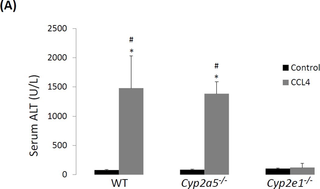
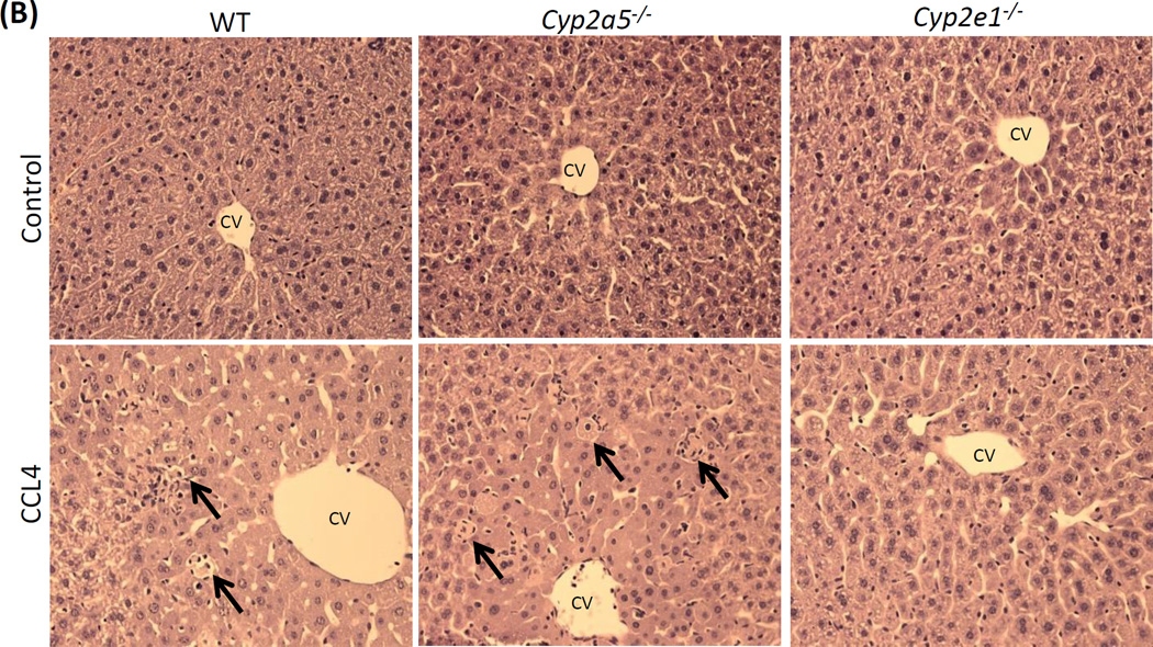
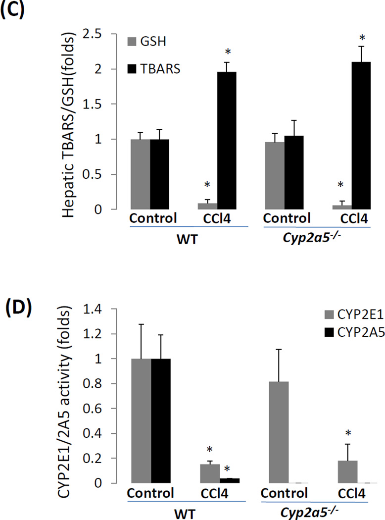
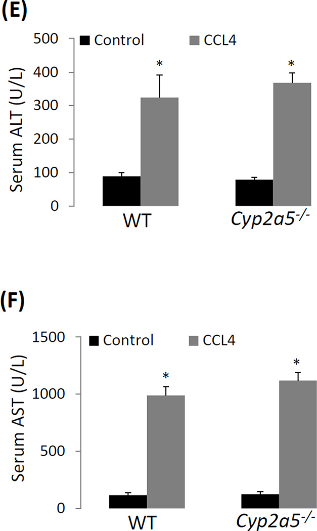
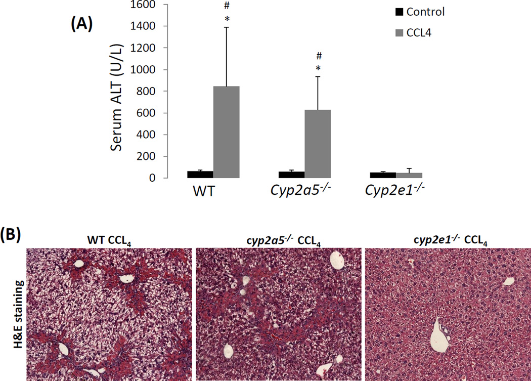
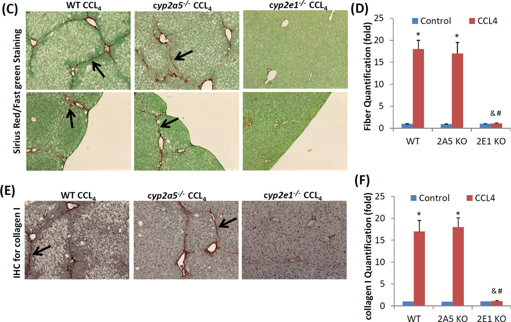
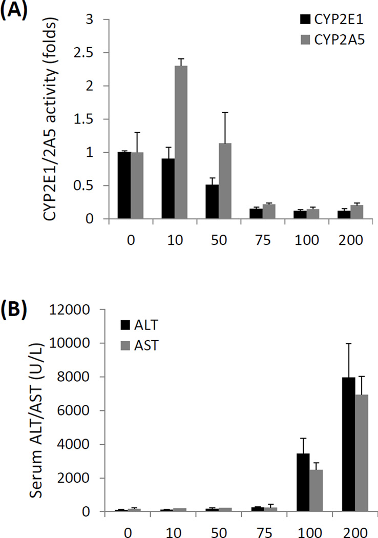
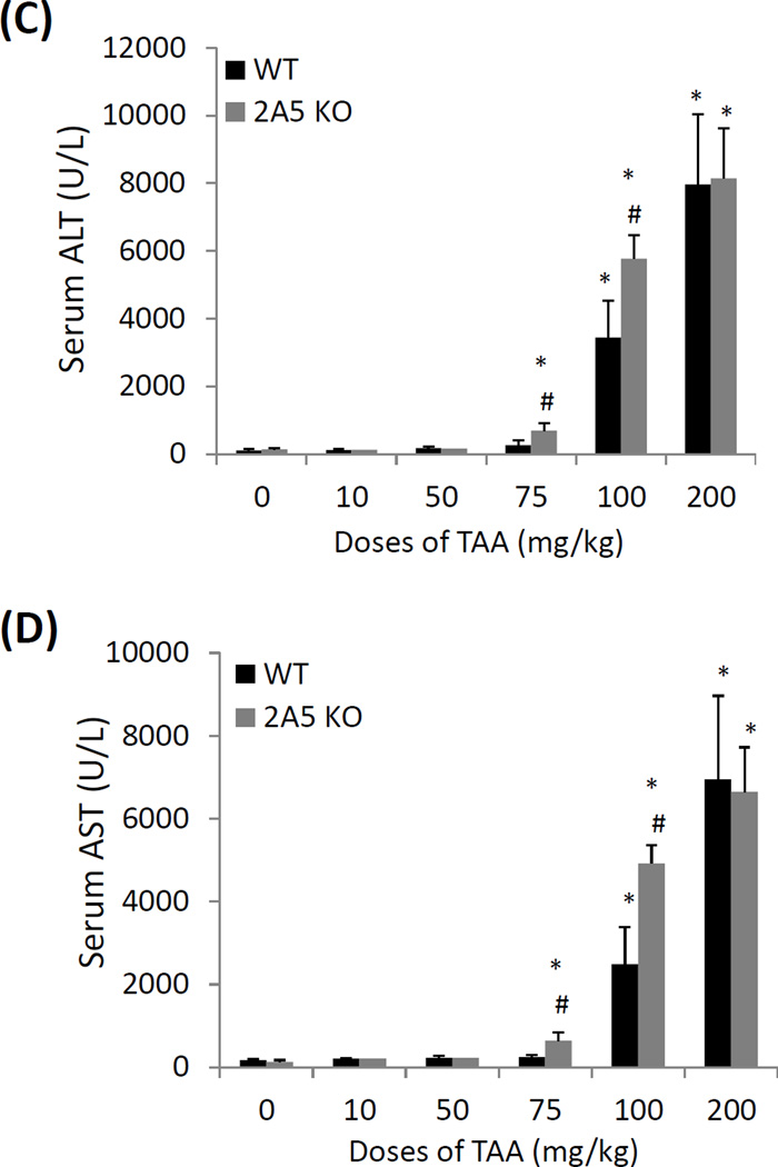
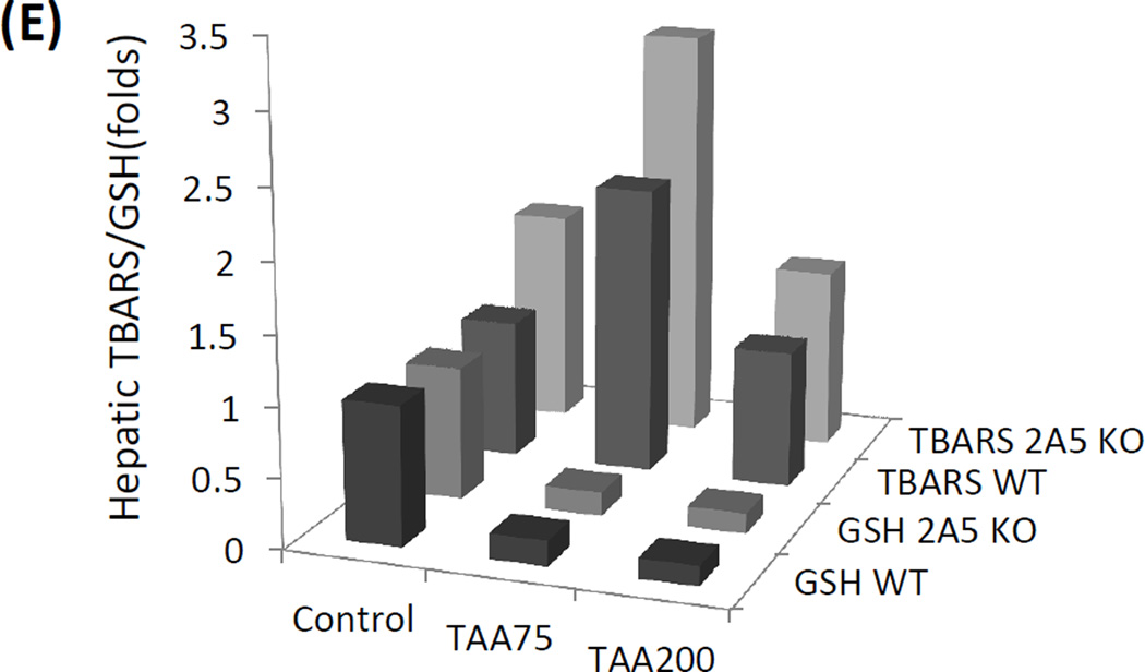
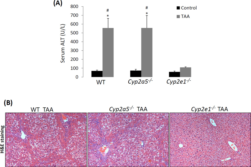
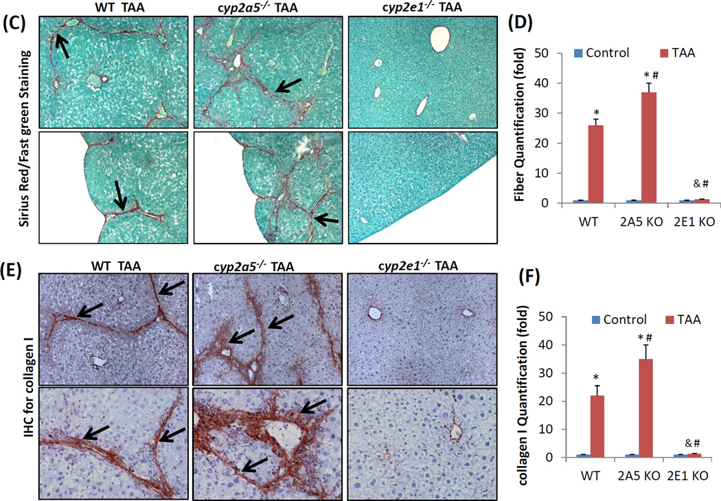
Liver injuries induced by carbon tetrachloride (CCL4) or thioacetamide (TAA) are dependent on cytochrome P450 2E1 (CYP2E1). CYP2A5 can be induced by TAA but not by CCL4. In this study, liver injury including fibrosis induced by CCL4 or TAA were investigated in wild-type (WT) mice and CYP2A5 knockout (cyp2a5 (-/-) ) mice as well as in CYP2E1 knockout (cyp2e1 (-/-) ) mice as a comparison. Acute and subchronic liver injuries including fibrosis were induced by CCL4 and TAA in WT mice but not in cyp2e1 (-/-) mice, confirming the indispensable role of CYP2E1 in CCL4 and TAA hepatotoxicity. WT mice and cyp2a5 (-/-) mice developed comparable acute liver injury induced by a single injection of CCL4 as well as subchronic liver injury including fibrosis induced by 1 month of repeated administration of CCL4, suggesting that CYP2A5 does not affect CCL4-induced liver injury and fibrosis. However, while 200 mg/kg TAA-induced acute liver injury was comparable in WT mice and cyp2a5 (-/-) mice, 75 and 100 mg/kg TAA-induced liver injury were more severe in cyp2a5 (-/-) mice than those found in WT mice. After multiple injections with 200 mg/kg TAA for 1 month, while subchronic liver injury as indicated by serum aminotransferases was comparable in WT mice and cyp2a5 (-/-) mice, liver fibrosis was more severe in cyp2a5 (-/-) mice than that found in WT mice. These results suggest that while both CCL4- and TAA-induced liver injuries and fibrosis are CYP2E1 dependent, under some conditions, CYP2A5 may protect against TAA-induced liver injury and fibrosis, but it does not affect CCL4 hepatotoxicity.

Keywords: Cytochrome P450; Fibrosis; Hepatic stellate cell; Liver injury; Metabolism; Oxidative stress.


Conflict of interest statement

The authors declare that there are no conflicts of interest.

Figures

Figure 1
Figure 1
CCL4-induced liver injury and oxidative stress are CYP2E1 dependent but independent of CYP2A5. (A) Serum ALT activity and (B) pathological observation in liver sections with H&E staining (×200) after CCL4 was injected i.p. at 0.5 ml/kg into WT, cyp2a5−/− mice and cyp2e1−/− mice. CV, central vein. Arrows show small single necrotic foci. (C) Liver TBARS and GSH and (D) liver microsomal CYP2E1 and CYP2A5 activity after CCL4 was injected i.p. at 0.5 ml/kg into WT and cyp2a5−/− mice. (E) Serum ALT activity and (F) Serum AST activity after CCL4 was injected i.p. at 15 µl/kg into WT and cyp2a5−/− mice. * P<0.05, compared with Control; # P<0.05, compared with cyp2e1−/− mice group.
Figure 1
Figure 1
CCL4-induced liver injury and oxidative stress are CYP2E1 dependent but independent of CYP2A5. (A) Serum ALT activity and (B) pathological observation in liver sections with H&E staining (×200) after CCL4 was injected i.p. at 0.5 ml/kg into WT, cyp2a5−/− mice and cyp2e1−/− mice. CV, central vein. Arrows show small single necrotic foci. (C) Liver TBARS and GSH and (D) liver microsomal CYP2E1 and CYP2A5 activity after CCL4 was injected i.p. at 0.5 ml/kg into WT and cyp2a5−/− mice. (E) Serum ALT activity and (F) Serum AST activity after CCL4 was injected i.p. at 15 µl/kg into WT and cyp2a5−/− mice. * P<0.05, compared with Control; # P<0.05, compared with cyp2e1−/− mice group.
Figure 2
Figure 2
CCL4-induced chronic liver injury and fibrosis are dependent on CYP2E1 but independent of CYP2A5. CCL4 was injected i.p. at 0.5 ml/kg into WT, cyp2a5−/− mice and cyp2e1−/− mice, twice a week, for one month. The mice were sacrificed 48 h after the last CCL4 injection. (A) Serum ALT activity. (B) H&E staining (×100). (C) Sirius Red/Fast Green staining(×100). Lower panels show the coarse surfaces of liver. Arrows show red stained fibers. (D) Quantification for Sirius Red staining. (E) IHC for collagen I (×100). Arrows show red stained collagen I. (F) Quantification for Collagen I IHC staining. * P<0.05, compared with Control; # P<0.05, compared with WT group; & P<0.05, compared with cyp2a5−/− mice group.
Figure 2
Figure 2
CCL4-induced chronic liver injury and fibrosis are dependent on CYP2E1 but independent of CYP2A5. CCL4 was injected i.p. at 0.5 ml/kg into WT, cyp2a5−/− mice and cyp2e1−/− mice, twice a week, for one month. The mice were sacrificed 48 h after the last CCL4 injection. (A) Serum ALT activity. (B) H&E staining (×100). (C) Sirius Red/Fast Green staining(×100). Lower panels show the coarse surfaces of liver. Arrows show red stained fibers. (D) Quantification for Sirius Red staining. (E) IHC for collagen I (×100). Arrows show red stained collagen I. (F) Quantification for Collagen I IHC staining. * P<0.05, compared with Control; # P<0.05, compared with WT group; & P<0.05, compared with cyp2a5−/− mice group.
Figure 3
Figure 3
Relationship of CYP2A5 and TAA-induced liver injury and oxidative stress. (A) Liver microsomal activities of CYP2E1 and CYP2A5 and (B) serum levels of ALT and AST after the WT mice were injected with different doses of TAA. (C) Serum levels of ALT, (D) serum AST and (E) hepatic TBARS and GSH after different doses of TAA were injected into WT mice and cyp2a5−/− mice. * P<0.05, compared with Control group (0 mg/kg); # P<0.05, compared with WT group.
Figure 3
Figure 3
Relationship of CYP2A5 and TAA-induced liver injury and oxidative stress. (A) Liver microsomal activities of CYP2E1 and CYP2A5 and (B) serum levels of ALT and AST after the WT mice were injected with different doses of TAA. (C) Serum levels of ALT, (D) serum AST and (E) hepatic TBARS and GSH after different doses of TAA were injected into WT mice and cyp2a5−/− mice. * P<0.05, compared with Control group (0 mg/kg); # P<0.05, compared with WT group.
Figure 4
Figure 4
TAA-induced liver fibrosis but not chronic liver injury is enhanced in the cyp2a5−/− mice compared to WT mice. TAA was injected at 200 mg/kg into WT mice, cyp2a5−/− mice and cyp2e1−/− mice, twice a week, for one month. The mice were sacrificed 48 h after the last TAA injection. (A) Serum ALT activity. (B) H&E staining (×100). (C) Sirius Red/Fast Green staining(×100). Lower panels show the coarse surfaces of liver. Arrows show red stained fibers. (D) Quantification for Sirius Red staining. (E) IHC for collagen I (×100 upper panels; ×200 lower panels). Arrows show red stained collagen I. (F) Quantification for Collagen I IHC staining. * P<0.05, compared with Control; # P<0.05, compared with WT group; & P<0.05, compared with cyp2a5−/− mice group.
Figure 4
Figure 4
TAA-induced liver fibrosis but not chronic liver injury is enhanced in the cyp2a5−/− mice compared to WT mice. TAA was injected at 200 mg/kg into WT mice, cyp2a5−/− mice and cyp2e1−/− mice, twice a week, for one month. The mice were sacrificed 48 h after the last TAA injection. (A) Serum ALT activity. (B) H&E staining (×100). (C) Sirius Red/Fast Green staining(×100). Lower panels show the coarse surfaces of liver. Arrows show red stained fibers. (D) Quantification for Sirius Red staining. (E) IHC for collagen I (×100 upper panels; ×200 lower panels). Arrows show red stained collagen I. (F) Quantification for Collagen I IHC staining. * P<0.05, compared with Control; # P<0.05, compared with WT group; & P<0.05, compared with cyp2a5−/− mice group.

Similar articles

Cited by

References

    1. Abu-Bakar A, Satarug S, Marks GC, Lang MA, Moore MR. Acute cadmium chloride administration induces hepatic and renal CYP2A5 mRNA, protein and activity in the mouse: involvement of transcription factor NRF2. Toxicol Lett. 2004;148:199–210. - PubMed
    1. Abu-Bakar A, Hakkola J, Juvonen R, Rahnasto-Rilla M, Raunio H, Lang MA. Function and regulation of the Cyp2a5/CYP2A6 genes in response to toxic insults in the liver. Curr. Drug Metab. 2013;14:137–150. - PubMed
    1. Avasarala S, Yang L, Sun Y, Leung AW, Chan WY, Cheung WT, Lee SS. A temporal study on the histopathological, biochemical and molecular responses of CCl(4)-induced hepatotoxicity in Cyp2e1-null mice. Toxicology. 2006;228:310–322. - PubMed
    1. Brattin WJ, Glende EA, Jr, Recknagel RO. Pathological mechanisms in carbon tetrachloride hepatotoxicity. J Free Radic Biol Med. 1985;1:27–38. - PubMed
    1. Cederbaum A. Nrf2 and antioxidant defense against CYP2E1 toxicity. Expert Opin. Drug Metab. Toxicol. 2009;5:1223–1244. - PubMed

Publication types

MeSH terms

LinkOut - more resources