. 2015 Oct;130(4):487-99.
doi: 10.1007/s00401-015-1477-1. Epub 2015 Sep 12.

Physiological amyloid-beta clearance in the periphery and its therapeutic potential for Alzheimer's disease

Affiliations

Physiological amyloid-beta clearance in the periphery and its therapeutic potential for Alzheimer's disease

Yang Xiang et al. Acta Neuropathol. 2015 Oct.

Abstract

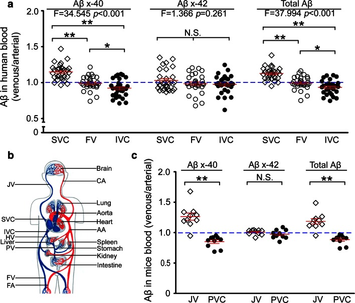
Amyloid-beta (Aβ) plays a pivotal role in the pathogenesis of Alzheimer's disease (AD). The physiological capacity of peripheral tissues and organs in clearing brain-derived Aβ and its therapeutic potential for AD remains largely unknown. Here, we measured blood Aβ levels in different locations of the circulation in humans and mice, and used a parabiosis model to investigate the effect of peripheral Aβ catabolism on AD pathogenesis. We found that blood Aβ levels in the inferior/posterior vena cava were lower than that in the superior vena cava in both humans and mice. In addition, injected (125)I labeled Aβ40 was located mostly in the liver, kidney, gastrointestinal tract, and skin but very little in the brain; suggesting that Aβ derived from the brain can be cleared in the periphery. Parabiosis before and after Aβ deposition in the brain significantly reduced brain Aβ burden without alterations in the expression of amyloid precursor protein, Aβ generating and degrading enzymes, Aβ transport receptors, and AD-type pathologies including hyperphosphorylated tau, neuroinflammation, as well as neuronal degeneration and loss in the brains of parabiotic AD mice. Our study revealed that the peripheral system is potent in clearing brain Aβ and preventing AD pathogenesis. The present work suggests that peripheral Aβ clearance is a valid therapeutic approach for AD, and implies that deficits in the Aβ clearance in the periphery might also contribute to AD pathogenesis.

Keywords: Alzheimer’s disease; Amyloid-beta; Clearance; Kidney; Liver; Parabiosis; Periphery.


Figures

Fig. 1
Fig. 1
Aβ concentrations at different locations of the systemic circulation. a The venous/arterial (V/A) ratio of Aβ concentrations among blood samples from different venous locations in humans (n = 30). The blue dotted line represents Aβ concentration in the femoral artery (FA) as a reference. b A diagram of the circulation system and sampling locations for Aβ measurement. The superior vena cava (SVC) collects blood from the head containing brain-derived Aβ. The inferior vena cava (IVC) proximal to the hepatic vein (HV) collects blood from the lower part of the body including liver, kidney, and gastrointestinal tract. The femoral vein (FV) contains metabolites after the circulation through the lower limbs. The femoral artery (FA) contains blood virtually identical to that in the aorta and is used as a reference. c The venous/arterial (V/A) ratio of Aβ concentrations among blood samples from different locations in APPswe/PS1 mice (n = 10). The blue dotted line represents the Aβ concentration in abdominal aorta (AA) as a reference. CA carotid artery, PV portal vein, JV jugular vein, SVC superior vena cava, IVC inferior vena cava, HV hepatic vein, FV femoral vein, FA femoral artery, PVC posterior vena cava. Mean ± SEM, one-way ANOVA and Tukey’s test for human plasma and 2-tailed t test for mouse plasma, *P < 0.05, **P < 0.01. N.S. no statistical significance
Fig. 2
Fig. 2
Parabiosis reduces brain amyloid burden of AD mice. a, c Representative images of Congo red and 6E10 immunohistochemical staining in neocortex and hippocampus in 9mon Tg and pa(3-9mon) Tg mice. Insets show the representative morphology at higher magnification. Scale bars 500 μm. b, d Comparison of the area fraction and density of Congo red or 6E10-positive Aβ plaques in the neocortex (Neoco.) and hippocampus (Hippo.) between 9mon Tg and pa(3-9mon) Tg mice. e, f, g Comparison of Aβ40, Aβ42 and total Aβ levels measured with ELISA in TBS, 2 % SDS and 70 % formic acid fractions of brain extracts between 9mon Tg and pa(3-9mon) Tg mice. h Illustration of Cerebral amyloid angiopathy (CAA) by immunofluorescence with the antibody to Aβ (6E10) and smooth muscle in the vessel wall (1A4). The arrow indicates the Aβ plaques in the brain parenchyma near the CAA. Scale bars 100 μm. i CAA visualized using Congo red staining. Insets show the representative morphology of CAA stained by Congo red at higher magnification. Scale bars 500 μm. j Comparison of numbers of CAA profiles and area fraction of CAA between 9mon Tg and pa(3-9mon) Tg mice. n = 8 per group, mean ± SEM, 2-tailed t test, *P < 0.05, **P < 0.01
Fig. 3
Fig. 3
Parabiosis attenuates neuroinflammation and Tau phosphorylation. a Representative images of astrocytosis stained with anti-GFAP antibody in the brain. Insets show the representative morphology at higher magnification. Scale bars 500 μm. b Immunofluorescence image of amyloid deposition and astrocytosis co-stained with 6E10 (green) and anti-GFAP (red) antibodies. Aβ plaques were surrounded by activated astrocytes. Scale bars 100 μm. c Representative images of microgliosis stained with anti-CD45 antibody in the brain. Insets show the representative morphology at higher magnification. Scale bars 500 μm. d Immunofluorescence image of amyloid deposition and microgliosis co-stained with 6E10 and anti-CD45 antibodies. Aβ plaques were surrounded by activated microglia. Scale bars 50 μm. e, f Comparisons of area fraction and cell density of astrocytosis (e) and microgliosis (f) in the neocortex (Neoco.) and hippocampus (Hippo.) among pa(3-9mon)Tg mice, control Tg mice and Wt mice. g Representative images of intracellular Tau phosphorylation stained with anti-pSer396 antibody in the brain. Insets show the representative morphology at higher magnification. Scale bars 500 μm. h Western blot assays of phosphorylated Tau at multiple sites including pSer199, pSer396, and total Tau (Tau5) in the brain homogenates of parabiotic Tg mice (PaTg), control Tg mice and wild-type mice (Wt). i Comparisons of area fraction and cell density of cells containing phosphorylated Tau stained with anti-pSer396 antibody in the neocortex among parabiotic Tg mice (PaTg), control Tg mice and wild-type mice. j Comparison of band density for phosphorylated Tau (pS199 and pS396) and total Tau (Tau5) shown in h among pa(3-9mon)Tg mice, control Tg mice and Wt mice. n = 8 per group, mean ± SEM., one-way ANOVA and Tukey’s test, *P < 0.05, **P < 0.01. N.S. no statistical significance
Fig. 4
Fig. 4
Parabiosis alleviates neuronal degeneration and loss in the hippocampus of pa(3-9mon)Tg mice. a Representative images of neurons and dendrites at CA1 region of hippocampus stained with anti-NeuN and anti-MAP-2 immunofluorescence in pa(3-9mon)Tg mice, control Tg mice, and wild-type (Wt) mice. Scale bars 100 μm. b Comparison of the area fractions of NeuN and MAP-2 staining among pa(3-9mon)Tg mice, control Tg mice, and Wt mice. c Comparison of area fractions of caspase-3 staining among pa(3-9mon)Tg mice, control Tg mice, and Wt mice. d Representative images of neuronal apoptosis at CA3 region of hippocampus as stained with activated caspase-3 immunofluorescence. Scale bars 100 μm. e Western blot assays of synapse-associated proteins including PSD93, PSD95, synapsin1 (SYN-1), and synaptophysin (Synap.) in brain homogenates of pa(3-9mon)Tg mice, control Tg mice, and wild-type (Wt) mice. f Comparisons of band density of PSD93, PSD95, SYN-1, and Synap. among pa(3-9mon)Tg mice, control Tg mice, and wild-type mice. n = 8 per group, mean ± SEM, one-way ANOVA and Tukey’s test, *P < 0.05, **P < 0.01

References

    1. Ajami B, Bennett JL, Krieger C, Tetzlaff W, Rossi FM. Local self-renewal can sustain CNS microglia maintenance and function throughout adult life. Nat Neurosci. 2007;10:1538–1543. doi: 10.1038/nn2014. - DOI - PubMed
    1. Bradshaw EM, Chibnik LB, Keenan BT, Ottoboni L, Raj T, Tang A, Rosenkrantz LL, Imboywa S, Lee M, Von Korff A, Alzheimer Disease Neuroimaging I. Morris MC, Evans DA, Johnson K, Sperling RA, Schneider JA, Bennett DA, De Jager PL. CD33 Alzheimer’s disease locus: altered monocyte function and amyloid biology. Nat Neurosci. 2013;16:848–850. doi: 10.1038/nn.3435. - DOI - PMC - PubMed
    1. Bu XL, Yao XQ, Jiao SS, Zeng F, Liu YH, Xiang Y, Liang CR, Wang QH, Wang X, Cao HY, Yi X, Deng B, Liu CH, Xu J, Zhang LL, Gao CY, Xu ZQ, Zhang M, Wang L, Tan XL, Xu X, Zhou HD, Wang YJ. A study on the association between infectious burden and Alzheimer’s disease. Eur J Neurol. 2014 - PubMed
    1. Conboy IM, Conboy MJ, Wagers AJ, Girma ER, Weissman IL, Rando TA. Rejuvenation of aged progenitor cells by exposure to a young systemic environment. Nature. 2005;433:760–764. doi: 10.1038/nature03260. - DOI - PubMed
    1. Deng J, Hou H, Giunta B, Mori T, Wang YJ, Fernandez F, Weggen S, Araki W, Obregon D, Tan J. Autoreactive-Abeta antibodies promote APP beta-secretase processing. J Neurochem. 2012;120:732–740. doi: 10.1111/j.1471-4159.2011.07629.x. - DOI - PMC - PubMed

Publication types

MeSH terms