. 2016 May;35(21):2746-55.
doi: 10.1038/onc.2015.340. Epub 2015 Sep 14.

LncRNA HOTAIR enhances ER signaling and confers tamoxifen resistance in breast cancer

Affiliations

LncRNA HOTAIR enhances ER signaling and confers tamoxifen resistance in breast cancer

X Xue et al. Oncogene. 2016 May.

Abstract

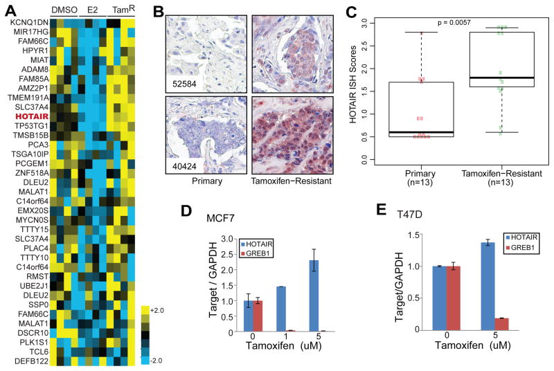
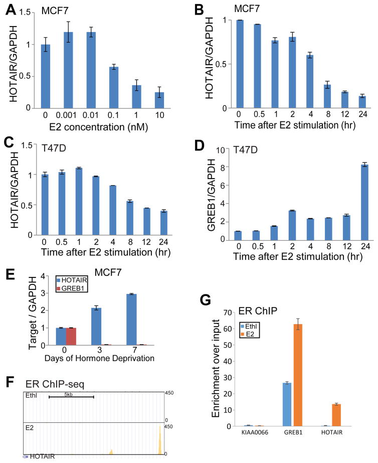
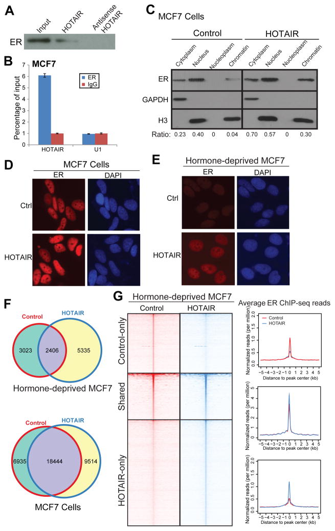
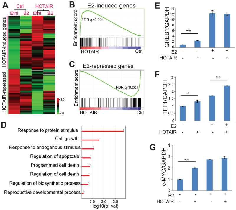
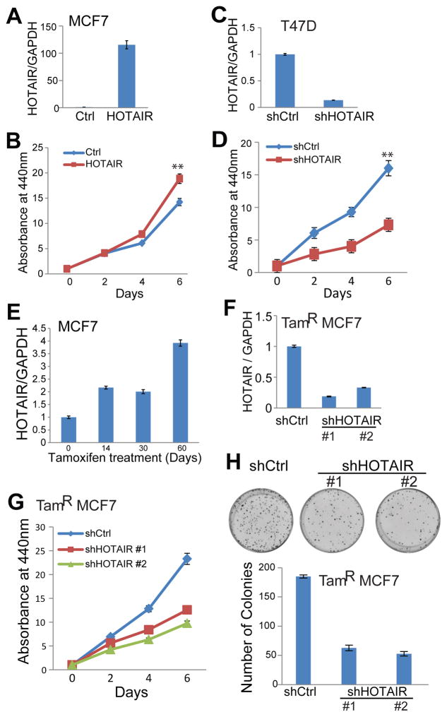
Tamoxifen, an estrogen receptor (ER) antagonist, is the mainstay treatment of breast cancer and the development of resistance represents a major obstacle for a cure. Although long non-coding RNAs such as HOTAIR have been implicated in breast tumorigenesis, their roles in chemotherapy resistance remain largely unknown. In this study, we report that HOTAIR (HOX antisense intergenic RNA) is upregulated in tamoxifen-resistant breast cancer tissues compared to their primary counterparts. Mechanistically, HOTAIR is a direct target of ER-mediated transcriptional repression and is thus restored upon the blockade of ER signaling, either by hormone deprivation or by tamoxifen treatment. Interestingly, this elevated HOTAIR increases ER protein level and thus enhances ER occupancy on the chromatin and potentiates its downstream gene regulation. HOTAIR overexpression is sufficient to activate the ER transcriptional program even under hormone-deprived conditions. Functionally, we found that HOTAIR overexpression increases breast cancer cell proliferation, whereas its depletion significantly impairs cell survival and abolishes tamoxifen-resistant cell growth. In conclusion, the long non-coding RNA HOTAIR is directly repressed by ER and its upregulation promotes ligand-independent ER activities and contributes to tamoxifen resistance.


Conflict of interest statement

Conflict of Interest

The authors declare no conflict of interest.

Figures

Figure 1
Figure 1. HOTAIR is up-regulated in tamoxifen-resistant breast cancer
A. Heatmap showing lncRNAs that are repressed by estradiol (E2) but up-regulated in tamoxifen-resistant (TamR) MCF7 cells. Microarray data were downloaded from GEO with GSE5840 and reanalyzed for lncRNA expression. HOTAIR is shown in red. B. HOTAIR ISH staining in 2 representative pairs of primary and tamoxifen-resistant breast cancers. C. Boxplot showing HOTAIR ISH staining intensity in a set (n=13) of matched primary and tamoxifen-resistant breast cancers. D–E. HOTAIR expression is increased by tamoxifen treatment. QRT-PCR analysis of HOTAIR and GREB1 was done in MCF7 (D) and T47D (E) cells treated with increasing doses of tamoxifen for 7 days. Gene expression was normalized to GAPDH. Data shown are mean ± SEM and is a representative of at least two independent experiments.
Figure 2
Figure 2. The lncRNA HOTAIR is directly repressed by estrogen through the ER
A. Estrogen inhibits HOTAIR expression in a dose-dependent manner. MCF7 cells were hormone starved for 3 days and treated with increasing amounts of estradiol (E2) for 6 hours. RNAs were then collected and subjected to qRT-PCR analysis of gene expression B. Estrogen inhibits HOTAIR expression in a time-dependent manner. MCF7 cells were treated with 1nM E2 and collected at different time-points for gene expression analysis by qRT-PCR. C–D. Estrogen inhibits HOTAIR expression in T47D cells. T47D cells were hormone deprived for 3 days followed by E2 stimulation for up to 24 hours. Cells were then collected at different time-points for qRT-PCR analysis of HOTAIR and GREB1 expression and normalized to GAPDH. E. Estrogen depletion restores HOTAIR level. QRT-PCR analysis of HOTAIR and GREB1 in MCF7 cells subjected for hormone deprivation for up to 7 days. F. Genome Browser view of ER binding events at enhancerd of the HOTAIR gene. ER ChIP-seq was performed in MCF7 cells stimulated with ethanol (Ethl) or estradiol (E2) and re-analyzed using HOMER (GSE23893). G. ChIP-qPCR showing ER binding to the HOTAIR distal enhancer. ER ChIP was performed in hormone-starved MCF7 cells stimulated with ethanol (vehicle) or E2 for 45 min. Enrichment of ER at specific genomic regions including GREB1 and HOTAIR enhancers were evaluated by qPCR. The KIAA0066 gene was utilized as a negative control as previously described.
Figure 3
Figure 3. HOTAIR interacts with the ER protein and enhances ER genomic action
A. HOTAIR lncRNA interacts with the ER protein. RNA pulldown assay was performed in MCF7 cells using biotin-labeled HOTAIR RNA probe transcribed in vitro. The antisense HOTAIR probe was used as negative control. B. ER protein binds to HOTAIR lncRNA. MCF7 cells were subjected to RIP assay using an anti-ER antibody or IgG control. IP-enriched RNA was then analyzed by qRT-PCR. U1 RNA was utilized as a negative control. C. HOTAIR overexpression increases ER protein level. MCF7 cell lysates were separated into cytoplasm, nuclear, nucleoplasm and chromatin-bound fractions and were detected by western blot analysis. GAPDH and H3 were utilized as loading controls for cytoplasmic and nuclear/chromatin fractions, respectively. Quantification was done by measuring band intensity with ImageJ and normalizing to loading control. D–E. Ectopic overexpression of HOTAIR increases nuclear ER level. ER immunostaining was performed in control and HOTAIR-overexpressing MCF7 cells grown in the presence (D) and absence (E) of estrogen. F. Overlap of ER-binding sites detected by ChIP-seq in MCF7 cells with control or HOTAIR overexpression in the absence and presence of estrogen. G. Heatmap depicting ER ChIP-seq read intensity around (±5_kb) peak centers detected in control or HOTAIR-overexpressing MCF7 cells under hormone-starved condition. Average ER ChIP-seq read intensity around ER binding sites (±5 kb) is shown on the right.
Figure 4
Figure 4. HOTAIR overexpression enhances ER transcriptional program
A. HOTAIR-induced and repressed genes are respectively increased and decreased by estrogen. Expression microarray was utilized to profile gene expression in control and HOTAIR-overexpressing MCF7 cells that were hormone-deprived for 3 days followed by either ethanol or 1nM E2 treatment for 6 hours. Expression of genes induced or repressed by HOTAIR for at least 2-fold were clustered and visualized using heatmap. B–C. Estrogen-induced genes are significantly enriched for up-regulation by HOTAIR (B), whereas estrogen-repressed genes down-regulated by HOTAIR (C). GSEA was carried out to determine the enrichment of E2-induced and -repressed gene sets in the expression dataset comparing control and HOTAIR-overexpressing MCF7 cells. D. HOTAIR-induced genes are enriched for cell growth and response to protein stimulus. GO analysis was performed using 132 genes that are increased by HOTAIR by at least 2 fold. Shown here are representative GO terms that are significantly enriched (P<0.05). E–G. HOTAIR enhances ER-target gene expression. QRT-PCR analysis of representative ER-induced genes were performed in control and HOTAIR-overexpressign MCF7 cells stimulated with ethanol or estrogen. Data were normalized to GAPDH. Error bars: mean± SEM. * indicates p<0.05, and ** indicates p<0.01.
Figure 5
Figure 5. HOTAIR promotes breast cancer cell growth and tamoxifen resistance
A–B. HOTAIR overexpression increases breast cancer cell growth. HOTAIR was overexpressed in MCF7 cells through lentiviral transduction with overexpression confirmed by qRT-PCR (A). Cell proliferation was determined by WST1 cell growth assay (B). Data shown are mean ± SEM and is a representative of at least two independent experiments. ** indicates p<0.01.C–D. HOTAIR knockdown decreases breast cancer cell growth. HOTAIR was depleted in T47D cells through shRNA lentiviral transduction (C) and cell growth was evaluated by WST1 assay (D). Data shown are mean ± SEM and is a representative of at least two independent experiments. **indicates p<0.01 E. HOTAIR is up-regulated in tamoxifen-resistant breast cancer cells. MCF7 cells were continuously grown in medium containing 5uM tamoxifen and periodically harvested for RNA isolation and qRT-PCR analysis. F–G. HOTAIR knockdown decreases tamoxifen-resistant breast cancer cell growth. HOTAIR was depleted in tamoxifen-resistant (TamR) MCF7 cells through lentiviral transduction of two shRNA constructs. Gene expression was determined by qRT-PCR (F) and cell growth by WST1 assay (G). H. HOTAIR knockdown inhibits colony formation abilities of breast cancer cells. Colony formation assays and quantifications were performed in tamoxifen-resistant MCF7 cells stably expressing control or HOTAIR-targeting shRNAs.

References

    1. Bussemakers MJ, van Bokhoven A, Verhaegh GW, Smit FP, Karthaus HF, Schalken JA, et al. DD3: a new prostate-specific gene, highly overexpressed in prostate cancer. Cancer research. 1999;59:5975–5979. - PubMed
    1. Chen D, Sun Y, Wei Y, Zhang P, Rezaeian AH, Teruya-Feldstein J, et al. LIFR is a breast cancer metastasis suppressor upstream of the Hippo-YAP pathway and a prognostic marker. Nature medicine. 2012;18:1511–1517. - PMC - PubMed
    1. Chisholm KM, Wan Y, Li R, Montgomery KD, Chang HY, West RB. Detection of long non-coding RNA in archival tissue: correlation with polycomb protein expression in primary and metastatic breast carcinoma. PloS one. 2012;7:e47998. - PMC - PubMed
    1. Clarke R, Leonessa F, Welch JN, Skaar TC. Cellular and molecular pharmacology of antiestrogen action and resistance. Pharmacological reviews. 2001;53:25–71. - PubMed
    1. Davis ME, Zuckerman JE, Choi CH, Seligson D, Tolcher A, Alabi CA, et al. Evidence of RNAi in humans from systemically administered siRNA via targeted nanoparticles. Nature. 2010;464:1067–1070. - PMC - PubMed

Publication types

MeSH terms