. 2015 Sep 28;34(6):642-55.
doi: 10.1016/j.devcel.2015.08.014. Epub 2015 Sep 10.

Identification of Happyhour/MAP4K as Alternative Hpo/Mst-like Kinases in the Hippo Kinase Cascade

Affiliations

Identification of Happyhour/MAP4K as Alternative Hpo/Mst-like Kinases in the Hippo Kinase Cascade

Yonggang Zheng et al. Dev Cell. .

Abstract

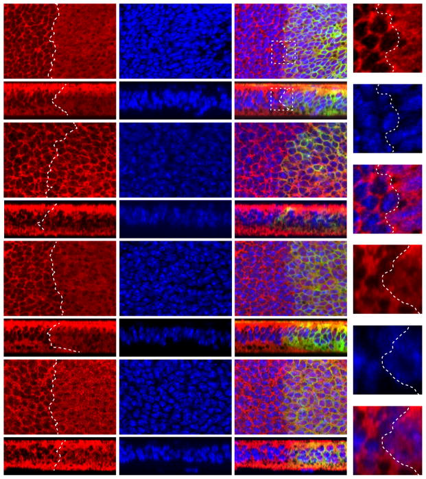
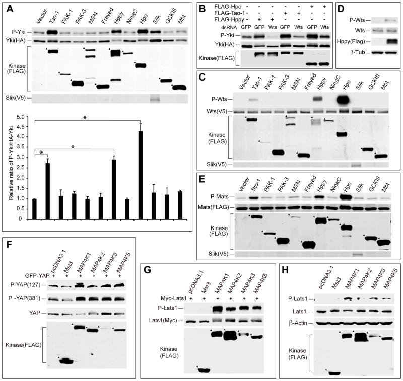
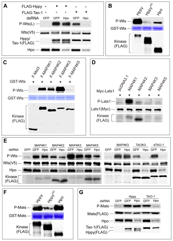
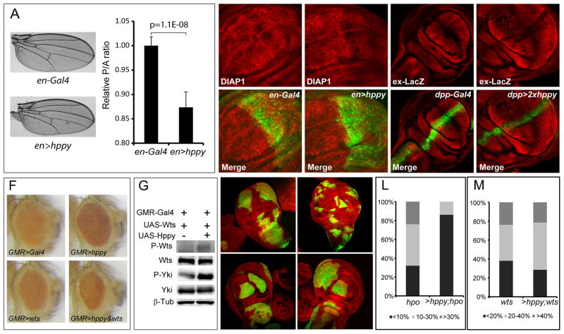
In Drosophila and mammals, the canonical Hippo kinase cascade is mediated by Hpo/Mst acting through the intermediary kinase Wts/Lats to phosphorylate the transcriptional coactivator Yki/YAP/TAZ. Despite recent reports linking Yki/YAP/TAZ activity to the actin cytoskeleton, the underlying mechanisms are poorly understood and/or controversial. Using Drosophila imaginal discs as an in vivo model, we show that Wts, but not Hpo, is genetically indispensable for cytoskeleton-mediated subcellular localization of Yki. Through a systematic screen, we identify the Ste-20 kinase Happyhour (Hppy) and its mammalian counterpart MAP4K1/2/3/5 as an alternative kinase that phosphorylates the hydrophobic motif of Wts/Lats in a similar manner as Hpo/Mst. Consistent with their redundant function as activating kinases of Wts/Lats, combined loss of Hpo/Mst and Hppy/MAP4K abolishes cytoskeleton-mediated regulation of Yki/YAP subcellular localization, as well as YAP cytoplasmic translocation induced by contact inhibition. These Hpo/Mst-like kinases provide an expanded view of the Hippo kinase cascade in development and physiology.


Figures

Figure 1
Figure 1. F-actin disruption induces nuclear exclusion of Yki in hpo but not wts mutant clones in Drosophila wing discs
Third instar larval wing discs containing GFP-positive mutant clones of the indicated genotypes were cultured in Schneider medium supplemented with (+LatB) or without (−LatB) 4μg/mL LatB for 1h and then stained with Yki antibody (Red) and DAPI (Blue). Both horizontal (A, E, G and I) and vertical (B, F, H and J) confocal sections are shown. Enlarged views of the boxed area in A″ and B″ are shown in C–C″ and D–D″, respectively. Without LatB treatment, both hpo and wts mutant clones showed clear nuclear staining of Yki (A, B, G and H). LatB treatment induced nuclear exclusion of Yki in hpo mutant clones (E and F) but not in wts mutant clones (I and J). See also Figure S1.
Figure 2
Figure 2. Identification of Hppy/MAP4K(1/2/3/5) as activators of Hippo signaling
(A) Eleven Drosophila Ste-20 kinases were individually expressed in S2R+ cells with HA-Yki. Cell lysates were probed for P-Yki S168, HA-Yki, FLAG-Ste20 kinases, or V5-Slik (upper panel). The relative ration of P-Yki versus total Yki was quantified (lower panel; mean±SD, n=3). Tao-1, Hppy, and Hpo increased the relative P-Yki/Yki ratio. Slik increased both Yki and P-Yki levels, but relative P-Yki/Yki ratio was not increased. * denotes a p-value<0.05. (B) wts RNAi abolished Yki phosphorylation induced by Hppy, Tao-1 or Hpo in S2R+ cells. Cell lysates expressing the indicated constructs were probed with the indicated antibodies. (C) Eleven Drosophila Ste-20 kinases were individually expressed in S2R+ cells with V5-Wts. Cell lysates were probed for P-Wts T1077, V5-Wts, FLAG-Ste20 like kinases, and V5-Slik. Tao-1, Hppy, and Hpo induced Wts phosphorylation. (D) Hppy promoted phosphorylation of endogenous Wts in S2R+ cells. Cell lysates expressing FLAG-Hppy were probed for P-Wts T1077. (E) Eleven Drosophila Ste-20 kinases were individually expressed in S2R+ cells with FLAG-Mats. Cell lysates were probed for P-Mob T12, FLAG-Mats, FLAG-Ste20 kinases, and V5-Slik. Tao-1, Hppy, and Hpo induced Mats phosphorylation. (F) 293T cells expressing the indicated constructs were probed for the phosphorylation of co-transfected GFP-YAP at YAPS127 and YAPS381 sites. (G) 293T cells expressing the indicated constructs were probed for the phosphorylation of co-transfected Myc-Lats1. (H) 293T cells expressing the indicated constructs were probed for the phosphorylation of endogenous Lats1. See also Figure S2.
Figure 3
Figure 3. Hppy/MAP4K(1/2/3/5) directly phosphorylates the hydrophobic motif of Wts/Lats
(A) S2R+ cells treated with the indicated dsRNAs were transfected with the indicated plasmids and probed for P-Wts, V5-Wts, FLAG-Tao-1, FLAG-Hppy, or endogenous Hpo. Note that hpo RNAi abolished Tao-1 induced but had little effect on Hppy induced Wts phosphorylation. (B) Hppy and Hpo phosphorylate Wts in vitro. Hppy, HppyKD, and Hpo expressed in S2R+ cells were purified by immunoprecipitation and incubated with GST-Wts, and the phosphorylation products were detected with P-Wts T1077 antibody. (C) In vitro kinase assay of MAP4K1/2/3/5. MAP4K1/2/3/5 expressed in 293T cells was purified by immunoprecipitation and incubated with GST-Wts, and the reaction products were probed for P-Wts. (D) MAP4K1 and MAP4K2 induce Lats1 phosphorylation in Mst1/2 null HCCs. MAP4K1, MAP4K2, MAP4K3, and MAP4K5 were individually expressed with Myc-Lats1 in Mst1/2 null HCCs, and followed by the analysis of Lats1 phosphorylation. (E) MAP4K1/2/3/5 and TAOK3 induces Wts phosphorylation in S2R+ cells. Note that hpo RNAi only slightly reduced Wts phosphorylation induced by MAP4K1/2/3/5 but abolished TAOK3 induced Wts phosphorylation. (F) Hppy and Hpo phosphorylate GST-Mats in vitro. Hppy, HppyKD, and Hpo expressed in S2R+ cells were purified by immunoprecipitation and incubated with GST-Mats, and the phosphorylation products were detected with P-Mob T12 antibody. (G) S2R+ cells treated with the indicated dsRNAs were transfected with the indicated plasmids and probed for P-Mob, FLAG-Mats, FLAG-Tao-1, or FLAG-Hppy. Note that hpo RNAi abolished Tao-1 induced but had little effects on Hppy induced Mats phosphorylation. See also Figure S3.
Figure 4
Figure 4. Hppy overexpression activates Hippo signaling in vivo and restricts growth of hpo but not wts mutant clones
(A) Hppy overexpression in the posterior compartment by en-Gal4 decreases wing size (left) and the relative ratio of posterior to anterior compartment (right; mean±SD, n=10). (B–C) Wing discs expressing GFP only (B) or GFP plus Hppy (C) by the en-Gal4 driver were stained for Diap1. Note the down-regulation of Diap1 expression in the poster compartment in the wing disc with Hppy overexpression (C). (D–E) Wing discs expressing GFP only (D) or GFP plus hppy (E) by the dpp-Gal4 driver were stained for the expression of ex-lacZ. Note the down-regulation of ex-lacZ expression upon hppy overexpression (arrows). The genotypes are: dpp-Gal4; UAS-GFP (D) and dpp-Gal4; UAS-GFP; UAS-hppy/UAS-hppy (E). (F) Hppy synergizes with Wts to reduce Drosophila eye size. Fly eyes from control (GMR-Gal4), Hppy-overexpression (GMR>hppy), Wts-overexpression (GMR>wts) or Hppy and Wts co-overexpression (GMR>hppy&wts) were imaged under the same magnification. Note that the fly eye overexpressing both Hppy and Wts is smaller than eyes overexpressing either Hppy or Wts. (G) Hppy promotes Wts and Yki phosphorylation in Drosophila eye tissues. Extracts made from fly heads overexpressing the indicated transgenes by the GMR-Gal4 driver were analyzed by western blotting. (H–K) Third instar larval wing discs containing GFP-positive clones of the indicated genotypes. hpo clones (H), hpo clones with Hppy overexpression (I), wts clones (J), wts clones with Hppy overexpression (K). Note that Hppy overexpression decreased the size of hpo clones. (L–M) Clone size distribution of the indicated genotypes. 20 discs were analyzed for each genotype. Clone size was quantified as the percentage of clone area relative to total wing area. Note that Hppy overexpression significantly decreased the size of hpo but not wts mutant clones.
Figure 5
Figure 5. Hppy functions redundantly with Hpo to regulate Yki in response to F-actin disruption
(A) Pupal eye discs containing GFP-negative hppy clones were stained with Ex antibody (Red). Clone boundary is marked by dotted line in (A′). Note the increased Ex expression in hppy mutant clone compared to the neighboring wildtype cells. (B–G) Third instar larval wing discs containing GFP-positive mutant clones of the indicated genotypes were cultured in Schneider medium supplemented with 4μg/mL LatB for 1h and then stained with Yki antibody (Red) and DAPI (Blue). Both horizontal (B, D, and F) and vertical (C, E, and G) confocal sections are shown. hppy;hpo mutant clone showed nuclear accumulation of Yki (B–C), and LatB treatment did not trigger Yki nuclear exclusion in hppy;hpo mutant clones (D–G). D–E and F–G represent two different hppy alleles recombined with hpo. (H) LatB-induced Yki phosphorylation in S2R+ cells was reduced by depletion of both hppy and hpo. S2R+ cells depleted with the indicated genes were treated with 1μg/mL LatB for 1 hr and then analyzed by western blotting (left). Relative P-Yki/Yki ratio was quantified in the graph (right; mean±SD, n=3)). Two non-overlapping dsRNAs against hppy were used (Hppy1 and Hppy2), alone or in combination with Hpo RNAi. * denotes a p-value<0.05. See also Figure S4.
Figure 6
Figure 6. MAP4K1/2/3/5 is required for Hpo/Mst-independent phosphorylation and nuclear exclusion of YAP induced by F-actin disruption
(A) Concomitant depletion of all four MAP4Ks leads to elevation in the expression levels of three Hpo pathway target genes ctgf, cyr61, and survivin in the Mst1/2 null HCCs. Total RNAs were prepared from HCCs transfected with the indicated siRNAs and the relative mRNA levels of indicated genes were measured by qPCR (mean±SD, n=3). Note the significant increase of gene expression when all four kinases were depleted. ** denotes a p-value<0.01. (B) Concomitant depletion of all four MAP4Ks promotes the proliferation of Mst1/2 null HCCs. (C) Depletion of MAP4K1/2/3/5 by RNAi reduces LatB-induced YAP and Lats phosphorylation in the Mst1/2 null HCCs. HCCs cultured on fibronectin-coated surface at low cell density were treated with 1μg/mL LatB for 1 hr and then analyzed by western blotting. Note that LatB induced YAP and Lats phosphorylation (compare lanes 1 and 2). Both inductions were diminished when four kinases were depleted (compare lanes 2 and 6). (D–E) Depletion of MAP4K1/2/3/5 by RNAi attenuates LatB-induced nuclear exclusion of YAP in the Mst1/2 null HCCs. HCCs transfected with control siRNAs or siRNAs against MAP4K1/2/3/5 were cultured on chamber slides coated with fibronectin at low cell density. Cell were treated with 1μg/mL LatB for 1 hr and then fixed and stained with YAP antibody, phalloidin for F-actin, and DAPI for cell nuclei (C). 100 cells were quantified for YAP localization and the results were shown in (D). See also Figure S5.
Figure 7
Figure 7. MAP4K1/2/3/5 is required for Hpo/Mst-independent nuclear exclusion of YAP induced by high cell density
Mst1/2 null HCCs cultured at low (upper two rows) or high (lower two rows) cell density were transfected with control or MAP4K1/2/3/5 siRNAs for 3 days, fixed, and stained with YAP antibody and DAPI. Both types of cells showed nuclear YAP signal at low density. At high density, cells with control RNAi showed cytoplasmic localization of YAP while cells with MAP4K1/2/3/5 depletion showed more nuclear localization of YAP. See also Figure S6.

References

    1. Aragona M, Panciera T, Manfrin A, Giulitti S, Michielin F, Elvassore N, Dupont S, Piccolo S. A mechanical checkpoint controls multicellular growth through YAP/TAZ regulation by actin-processing factors. Cell. 2013;154:1047–1059. - PubMed
    1. Barry ER, Camargo FD. The Hippo superhighway: signaling crossroads converging on the Hippo/Yap pathway in stem cells and development. Current opinion in cell biology. 2013;25:247–253. - PubMed
    1. Boggiano JC, Fehon RG. Growth control by committee: intercellular junctions, cell polarity, and the cytoskeleton regulate Hippo signaling. Developmental cell. 2012;22:695–702. - PMC - PubMed
    1. Boggiano JC, Vanderzalm PJ, Fehon RG. Tao-1 phosphorylates Hippo/MST kinases to regulate the Hippo-Salvador-Warts tumor suppressor pathway. Developmental cell. 2011;21:888–895. - PMC - PubMed
    1. Bryk B, Hahn K, Cohen SM, Teleman AA. MAP4K3 regulates body size and metabolism in Drosophila. Developmental biology. 2010;344:150–157. - PubMed

Publication types

MeSH terms

LinkOut - more resources