

MGH-USC Human Connectome Project datasets with ultra-high b-value diffusion MRI
- PMID: 26364861
- PMCID: PMC4651764
- DOI: 10.1016/j.neuroimage.2015.08.075
MGH-USC Human Connectome Project datasets with ultra-high b-value diffusion MRI
Abstract
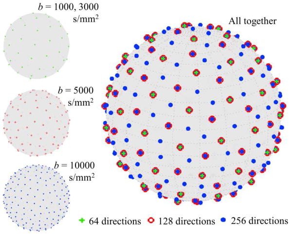
The MGH-USC CONNECTOM MRI scanner housed at the Massachusetts General Hospital (MGH) is a major hardware innovation of the Human Connectome Project (HCP). The 3T CONNECTOM scanner is capable of producing a magnetic field gradient of up to 300 mT/m strength for in vivo human brain imaging, which greatly shortens the time spent on diffusion encoding, and decreases the signal loss due to T2 decay. To demonstrate the capability of the novel gradient system, data of healthy adult participants were acquired for this MGH-USC Adult Diffusion Dataset (N=35), minimally preprocessed, and shared through the Laboratory of Neuro Imaging Image Data Archive (LONI IDA) and the WU-Minn Connectome Database (ConnectomeDB). Another purpose of sharing the data is to facilitate methodological studies of diffusion MRI (dMRI) analyses utilizing high diffusion contrast, which perhaps is not easily feasible with standard MR gradient system. In addition, acquisition of the MGH-Harvard-USC Lifespan Dataset is currently underway to include 120 healthy participants ranging from 8 to 90 years old, which will also be shared through LONI IDA and ConnectomeDB. Here we describe the efforts of the MGH-USC HCP consortium in acquiring and sharing the ultra-high b-value diffusion MRI data and provide a report on data preprocessing and access. We conclude with a demonstration of the example data, along with results of standard diffusion analyses, including q-ball Orientation Distribution Function (ODF) reconstruction and tractography.
Keywords: Adolescents; Children; Lifespan; Multi-shell HARDI; Older adults; Preprocessing.
Copyright © 2015 Elsevier Inc. All rights reserved.
Figures


References
-
- Anderson AW. Measurement of fiber orientation distributions using high angular resolution diffusion imaging. Magn Reson Med. 2005;54:1194–1206. - PubMed
-
- Andersson JL, Xu J, Yacoub E, Auerbach E, Moeller S, Ugurbil K. A comprehensive Gaussian Process framework for correcting distortions and movements in diffusion images. Proc Intl Soc Mag Reson Med. 2012:2426.
Publication types
MeSH terms
Grants and funding
- U01 MH093765/MH/NIMH NIH HHS/United States
- U54 EB020406/EB/NIBIB NIH HHS/United States
- P50 AG005134/AG/NIA NIH HHS/United States
- 1S10RR023401/RR/NCRR NIH HHS/United States
- K99 EB015445/EB/NIBIB NIH HHS/United States
- 1U54EB020406-01/EB/NIBIB NIH HHS/United States
- S10 RR023401/RR/NCRR NIH HHS/United States
- K99EB015445/EB/NIBIB NIH HHS/United States
- K01AG040197/AG/NIA NIH HHS/United States
- 5P41 EB015922-16/EB/NIBIB NIH HHS/United States
- P41 EB015922/EB/NIBIB NIH HHS/United States
- U01MH093765/MH/NIMH NIH HHS/United States
- S10 RR019307/RR/NCRR NIH HHS/United States
- R00 EB015445/EB/NIBIB NIH HHS/United States
- 1S10RR019307/RR/NCRR NIH HHS/United States
- 1S10RR023043/RR/NCRR NIH HHS/United States
- S10 RR023043/RR/NCRR NIH HHS/United States
- P50AG005134/AG/NIA NIH HHS/United States
- K01 AG040197/AG/NIA NIH HHS/United States
LinkOut - more resources
Full Text Sources
Other Literature Sources
Miscellaneous
