. 2015 Sep 14:5:14040.
doi: 10.1038/srep14040.

Experimental evaluation of the zoonotic infection potency of simian retrovirus type 4 using humanized mouse model

Affiliations

Experimental evaluation of the zoonotic infection potency of simian retrovirus type 4 using humanized mouse model

Kei Sato et al. Sci Rep. .

Abstract

During 2001-2002 and 2008-2011, two epidemic outbreaks of infectious hemorrhagic disease have been found in Japanese macaques (Macaca fuscata) in Kyoto University Primate Research Institute, Japan. Following investigations revealed that the causative agent was simian retrovirus type 4 (SRV-4). SRV-4 was isolated by using human cell lines, which indicates that human cells are potently susceptible to SRV-4 infection. These raise a possibility of zoonotic infection of pathogenic SRV-4 from Japanese macaques into humans. To explore the possibility of zoonotic infection of SRV-4 to humans, here we use a human hematopoietic stem cell-transplanted humanized mouse model. Eight out of the twelve SRV-4-inoculated humanized mice were infected with SRV-4. Importantly, 3 out of the 8 infected mice exhibited anemia and hemophagocytosis, and an infected mouse died. To address the possibility that SRV-4 adapts humanized mouse and acquires higher pathogenicity, the virus was isolated from an infected mice exhibited severe anemia was further inoculated into another 6 humanized mice. However, no infected mice exhibited any illness. Taken together, our findings demonstrate that the zoonotic SRV-4 infection from Japanese macaques to humans is technically possible under experimental condition. However, such zoonotic infection may not occur in the real society.


Figures
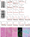
Figure 1
Figure 1. Dynamics of SRV-4 infection in humanized mouse model (experiment 1).
(a,b) Proviral DNA (a) and viral RNA (b) were respectively analyzed by PCR and RT-PCR (targeted to env region). ACTB and Actb (the murine ortholog of human ACTB) were used as internal control, and the RT-PCR without reverse transcriptase (–RT) was used as negative control. In a and b, the results of MLN in mouse no. 4 are not shown because MLN was not detected. (c-t) Longitudinal analyses on the dynamics of SRV-4 infection. X-axes indicate wpi. (c) The body weights were routinely measured and were shown as the ratio to the initial weight. (d-k) The numbers of white blood cells (d) red blood cells (e) and platelets (f) hematocrit (g) hemoglobin concentration (h) and the values of MCV (i), MCH (j) and MCHC (k) were measured by hematometry. (l-t) The numbers of human CD45+ leukocytes (l), CD3+ T cells (m) CD4+ T cells (CD3+CD4+ cells; (n) CD8+ T cells (CD3+CD8+ cells; (o) naïve CD4+ T cells (CD3+CD4+CD45RA+ cells; (p) memory CD4+ T cells (CD3+CD4+CD45RA cells; (q) naïve CD8+ T cells (CD3+CD8+CD45RA+ cells; (r) memory CD8+ T cells (CD3+CD8+CD45RA cells; (s) and CD19+ B cells (t) were analyzed by flow cytometry and hematometry. In c-t, red, an SRV-4-infected mouse exhibited anemia (no. 2); brown, an SRV-4-infected mouse (i.e., viral DNA was detected; no. 3); and black, SRV-inoculated mice without viral replication. The data from mock-infected mice (n = 3) is presented as averages with SEMs. The mouse numbers correspond to those in Table 1. (u-w) Hemophagocytosis in the liver of an SRV-4-infected mice exhibited anemia (no. 2). The liver section of the mouse was assessed by H&E staining (u, right), immunostaining (v), and Berlin blue staining (w), and the representatives are shown. In u, H&E staining of the liver section of mock-infected mouse is presented on the left panel. In u and v, areas enclosed with squares are enlarged in independent panels. Arrowheads indicate the degenerated hepatocytes. In w, the cells stained with blue indicate hemosiderin-containing hemophagocytic macrophages. CV, central vein; PV, portal vein. Scale bar, 100 μm. In a and b, gels have been cropped; full uncropped gels are available as Supplementary Figure 1.
Figure 2
Figure 2. Dynamics of SRV-4 infection in humanized mouse model (experiment 2).
(a) Proviral DNA was analyzed by PCR (targeted to env region). Actb/ACTB were used as internal control. In a, the results of thymus, MLN, and PB at 8 and 10 wpi in mouse no. 12 are not shown because this mouse was suddenly died at 6 wpi. (b-j) Longitudinal analyses on the dynamics of SRV-4 infection. X-axes indicate wpi. (b) The body weights were routinely measured and were shown as the ratio to the initial weight. (c-j) The numbers of white blood cells (c), red blood cells (d), platelets (e), hematocrit (f), hemoglobin concentration (g), and the values of MCV (h), MCH (i), and MCHC (j) were measured by hematometry. In b-j, red, SRV-4-infected mice exhibited anemia (no. 10 and 12); brown, SRV-4-infected mouse (i.e., viral DNA was detected; no. 11, 13-15); and black, an SRV-inoculated mouse without viral replication. Note that mouse no. 12 exhibited anemia and suddenly died at 6 wpi, which is indicated by daggers in b-j. The data from mock-infected mice (n = 3) were presented as the averages with SEMs. The mouse numbers correspond to those in Table 1. (k) Hemophagocytosis in the BM of an SRV-4-infected mice exhibited anemia (no. 10). The BM fluid smear was assessed by NSE staining, and a representative result is shown. Macrophages (brown) and neutrophils (blue) are stained. In a, gels have been cropped; full uncropped gels are available as Supplementary Figure 2.
Figure 3
Figure 3. The levels of human cytokines in plasma of SRV-4-infected humanized mice.
The amounts of IFN-γ, TNF-α, IL-6, IL-2, IL-4, and IL-10 in the plasma of mock-infected mice (n = 6), SRV-4-inoculated mice without infection (n = 4; black), SRV-4-infected mice without anemia (n = 5; brown), and SRV-4-infected mice exhibited anemia (n = 2; red) at 10 wpi are respectively shown. Horizontal bars indicate the averages of mock-infected mice (n = 6) and SRV-4-infected mice with or without anemia (n = 7), respectively.
Figure 4
Figure 4. Dynamics of SRV-4* infection in humanized mouse model (experiment 3).
(a-c) Virus isolation from infected humanized mice. (a) The human MNCs isolated from the BM and spleen of SRV-4-inoculated mice were cocultured with 293T cells. DNA was extracted from the cocultured 293T cells and proviral DNA was analyzed by PCR. (b,c) LacZ marker rescue assay. The detailed procedure is described in Methods, and representative results of LacZ marker rescue assay are shown in c. Arrowheads indicate the presence of blue foci. (d,e) Proviral DNA (d) and viral RNA (e) were respectively analyzed by PCR and RT-PCR (targeted to env region). Actb/ACTB were used as internal control, and–RT was used as negative control. (f-n) Longitudinal analyses on the dynamics of SRV-4* infection. X-axes indicate wpi. (f) The body weights were routinely measured and were shown as the ratio to the initial weight. (g-n) The numbers of white blood cells (g), red blood cells (h), platelets (i), hematocrit (j), hemoglobin concentration (k), and the values of MCV (l), MCH (m), and MCHC (n) were measured by hematometry. In f-n, brown, SRV-4-infected mice (i.e., viral DNA was detected; no. 19 and 21); and black, SRV-inoculated mice without viral replication. The data from mock-infected mice (n = 3) were presented as the averages with SEMs. The mouse numbers correspond to those in Table 1. In a, d, and e, gels have been cropped; full uncropped gels are available as Supplementary Figure 3.
Figure 5
Figure 5. Anti-SRV-4 ability of human APOBEC3 proteins.
(a) Dose-dependent anti-SRV-4 ability of human APOBEC3F and APOBEC3G. 293T cells were cotransfected with pSR415 (1 μg), pSRV4ψluc (1 μg), and flag-tagged APOBEC3G (blue) or APOBEC3F (red) expression plasmids (25, 50, 100, or 200 ng), and the infectivity of the released virions in the culture supernatant was measured as described in Methods. The infectivity is shown as the percentage of the value of APOBEC3-untransfected cells. (b,c) Western blotting. 293T cells were cotransfected with pSR415 (1 μg), pSRV4ψluc (1 μg), and flag-tagged APOBEC3G (blue) or APOBEC3F (red) expression plasmids (200 ng). The expression levels of viral proteins (Gag and CA) and flag-tagged APOBEC3s in the transfected cells, the amount of released virion in the culture supernatant (CA and TM), and the amount of APOBEC3 in the released virion was assessed by Western blotting. The input of cell lysate was standardized to α-tubulin (TUBA), and representative results are shown in b. The amount of APOBEC3s in the released virion was quantified as described in Methods, and the summarized data of the 6 independent experiments are shown in c. (d) Deaminase-dependent anti-SRV-4 ability of human APOBEC3F and APOBEC3G. 293T cells were cotransfected with pSR415 (1 μg), pSRV4ψluc (1 μg), and flag-tagged plasmid (200 ng) expressing wild-type (WT) APOBEC3G, APOBEC3G E259Q, WT APOBEC3F, or APOBEC3F E251Q. The infectivity of the released virions in the culture supernatant was measured as described in Methods and is shown as the percentage of the value of APOBEC3-untransfected cells. In a and d, the assays were performed in triplicate. The statistical difference is determined by Student’s t test, and statistically significant difference (P < 0.05) against no APOBEC3 is indicated by asterisks. Error bars represent SDs. In b, blots have been cropped; full uncropped blots are available as Supplementary Figure 4.

References

    1. Daniel M. D. et al.. A new type D retrovirus isolated from macaques with an immunodeficiency syndrome. Science 223, 602–605 (1984). - PubMed
    1. Marx P. A. et al.. Simian AIDS: isolation of a type D retrovirus and transmission of the disease. Science 223, 1083–1086 (1984). - PubMed
    1. Montiel N. A. An updated review of simian betaretrovirus (SRV) in macaque hosts. J. Med. Primatol. 39, 303–314 (2010). - PubMed
    1. Henrickson R. V. et al.. Clinical features of simian acquired immunodeficiency syndrome (SAIDS) in rhesus monkeys. Lab. Anim. Sci. 34, 140–145 (1984). - PubMed
    1. Committee on Disease Control, Primate Research Institute & University, K. Information of Hemorrhagic Syndrome of Japanese Macaques (Provisional Designation). Primate Research 26, 69–71 10.2354/psj.2326.2369 (2010). - DOI

Publication types

MeSH terms