. 2015 Oct 6;22(4):606-18.
doi: 10.1016/j.cmet.2015.08.018. Epub 2015 Sep 10.

MSC Transplantation Improves Osteopenia via Epigenetic Regulation of Notch Signaling in Lupus

Affiliations

MSC Transplantation Improves Osteopenia via Epigenetic Regulation of Notch Signaling in Lupus

Shiyu Liu et al. Cell Metab. .

Abstract

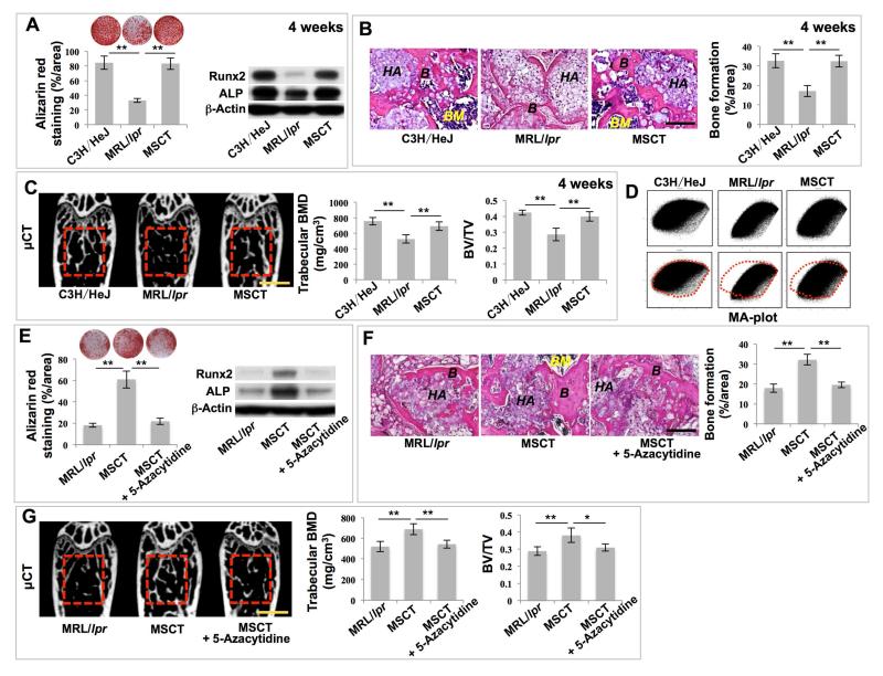
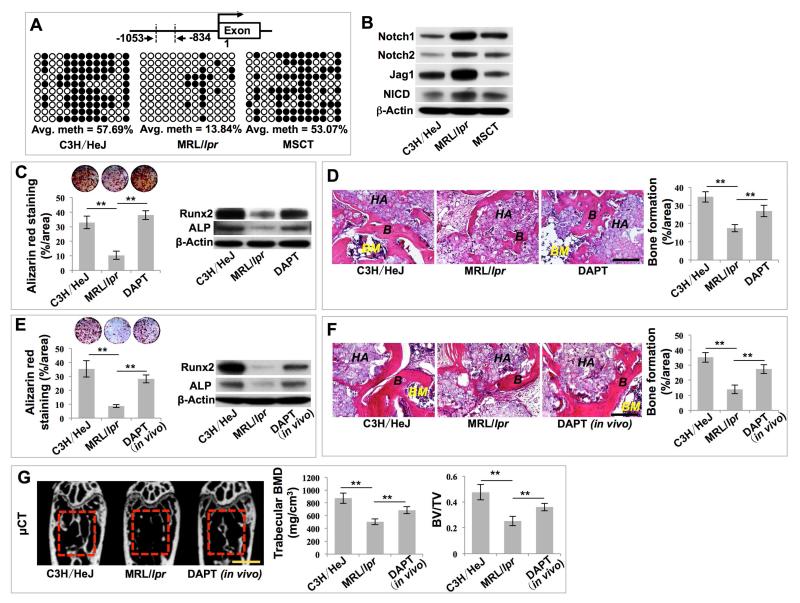
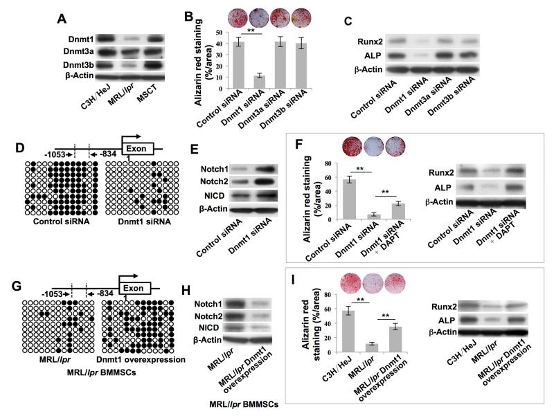
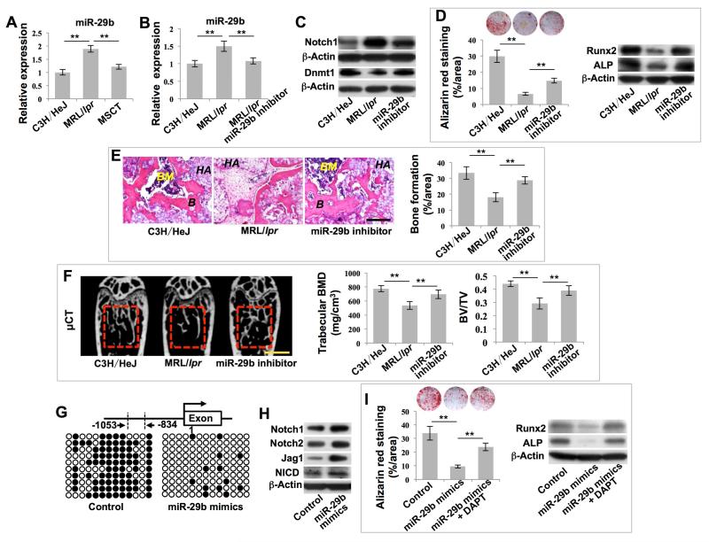
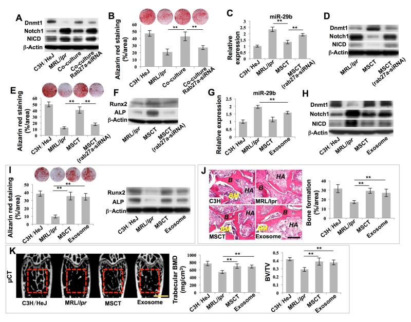
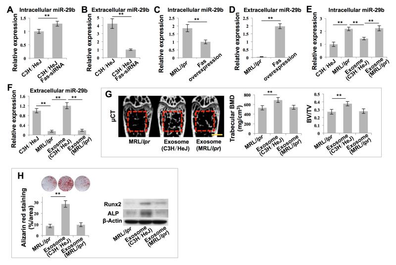
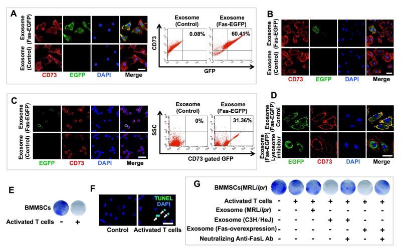
Mesenchymal stem cell transplantation (MSCT) has been used to treat human diseases, but the detailed mechanisms underlying its success are not fully understood. Here we show that MSCT rescues bone marrow MSC (BMMSC) function and ameliorates osteopenia in Fas-deficient-MRL/lpr mice. Mechanistically, we show that Fas deficiency causes failure of miR-29b release, thereby elevating intracellular miR-29b levels, and downregulates DNA methyltransferase 1 (Dnmt1) expression in MRL/lpr BMMSCs. This results in hypomethylation of the Notch1 promoter and activation of Notch signaling, in turn leading to impaired osteogenic differentiation. Furthermore, we show that exosomes, secreted due to MSCT, transfer Fas to recipient MRL/lpr BMMSCs to reduce intracellular levels of miR-29b, which results in recovery of Dnmt1-mediated Notch1 promoter hypomethylation and thereby improves MRL/lpr BMMSC function. Collectively our findings unravel the means by which MSCT rescues MRL/lpr BMMSC function through reuse of donor exosome-provided Fas to regulate the miR-29b/Dnmt1/Notch epigenetic cascade.


Figures

Figure 1
Figure 1. MSC transplantation (MSCT) rescued impaired BMMSC functions and osteoporotic phenotype in MRL/lpr mice via regulation of DNA methylation profile
(A) Alizarin red staining showed mineralized nodule formation of BMMSCs derived from wild-type C3H/HeJ mice (C3H/HeJ), MRL/lpr mice (MRL/lpr) and MRL/lpr mice at 4 weeks post-MSCT. n = 5. Western blot showed expression of Runx2 and ALP in BMMSCs. β-Actin was used as a loading control. (B) H&E staining showed formation of new bone (B) and bone marrow (BM) around HA/TCP (HA) carrier when BMMSCs were implanted into immunocompromised mice. n = 5. (C) μCT analysis of bone mineral density (BMD) and bone volume/total volume (BV/TV) of femurs. n = 5. (D) MA plot showed global methylation patterns of gene promoters in BMMSCs, as assessed by DNA methylation microarray. (E) Alizarin red staining showed mineralized nodule formation by BMMSCs. n = 5. Western blot showed expressions of Runx2 and ALP in BMMSCs. (F) H&E staining showed formation of new bone (B) and bone marrow (BM) around HA/TCP (HA) carrier when BMMSCs were implanted into immunocompromised mice. n = 5. (G) μCT analysis of BMD and BV/TV of femurs. n = 5. All results are representative of data generated in three independent experiments except DNA methylation microarray analysis. Statistical significance was determined with one-way analysis of variance (ANOVA). **P < 0.01; *P < 0.05. Error bars: mean ± SD; 200 μm (B, F), 1 mm (C, G).
Figure 2
Figure 2. MSCT inhibited Notch signaling in recipient MRL/lpr BMMSCs via regulating DNA methylation in Notch1 promoter region
(A) Bisulfite genomic sequencing analysis of BMMSC Notch1 promoter region. Each box is representative of the indicated BMMSC sample; each row of dots is representative of the CpG island in the Notch1 promoter region; each dot is representative of a single CpG. Empty dots indicate unmethylated CpGs; black dots indicate methylated CpGs. Each row represents a single sequenced clone (ten for each sample). (B) Western blot showed Notch1, Notch2, Jag1 and NICD expression in BMMSCs. (C) Alizarin red staining showed mineralized nodule formation by BMMSCs. n = 5. Western blot showed expressions of Runx2 and ALP in BMMSCs. (D) H&E staining showed formation of new bone (B) and bone marrow (BM) around HA/TCP (HA) carrier when BMMSCs were implanted into immunocompromised mice. n = 5. (E) Alizarin red staining showed mineralized nodule formation by BMMSCs. n = 5. Western blot showed expression of Runx2 and ALP in BMMSCs. (F) H&E staining showed formation of new bone (B) and bone marrow (BM) around HA/TCP (HA) carrier when BMMSCs were implanted into immunocompromised mice. n = 5. (G) μCT analysis of BMD and BV/TV of femurs. n = 5. All results are representative of data generated in three independent experiments. Statistical significance was determined with one-way ANOVA. **P < 0.01. Error bars: mean ± SD, 200 μm (D, F), 1 mm (G).
Figure 3
Figure 3. MSCT rescued hypomethylation of the Notch gene promoter in recipient MRL/lpr BMMSCs via upregulation of Dnmt1
(A) Western blot showed Dnmt1, Dnmt3a and Dnmt3b expression in BMMSCs. (B) Alizarin red staining showed mineralized nodule formation by BMMSCs. n = 4. (C) Western blot showed Runx2 and ALP expression in BMMSCs. (D, G) Bisulfite genomic sequencing analysis of BMMSC Notch1 promoter region. (E, H) Western blot showed Notch1, Notch2 and NICD expression in BMMSCs. (F, I) Alizarin red staining showed mineralized nodule formation by BMMSCs. n = 4. Western blot showed Runx2 and ALP expression in BMMSCs. All results are representative of data generated in three independent experiments. Statistical significance was determined with one-way ANOVA. **P < 0.01. Error bars: mean ± SD.
Figure 4
Figure 4. MSCT governed Dnmt1-mediated DNA methylation of the Notch1 promoter via regulating intracellular levels of miR-29b
(A, B) Real-time PCR showed miR-29b expression in BMMSCs. n = 5. (C) Western blot showed Notch1 and Dnmt1 expression in BMMSCs. (D) Alizarin red staining showed mineralized nodule formation by BMMSCs. n = 5. Western blot showed Runx2 and ALP expression in BMMSCs. (E) H&E staining showed formation of new bone (B) and bone marrow (BM) around HA/TCP (HA) carrier when BMMSCs were implanted into immunocompromised mice. n = 5. (F) μCT analysis showed BMD and BV/TV of femurs. n = 5. (G) Bisulfite genomic sequencing analysis of BMMSC Notch1 promoter region. (H) Western blot showed Notch1, Notch2, Jag1 and NICD expression in BMMSCs. (I) Alizarin red staining showed mineralized nodule formation by BMMSCs. n = 4. Western blot showed Runx2 and ALP expression in BMMSCs. All results are representative of data generated in three independent experiments. Statistical significance was determined with one-way ANOVA. **P < 0.01. Error bars: mean ± SD, 200 μm (E), 1 mm (F).
Figure 5
Figure 5. Exosomes secreted due to MSCT downregulated intracellular levels of miR-29b in recipient MRL/lpr BMMSCs
(A) Western blot showed Dnmt1, Notch1 and NICD expression in BMMSCs. (B) Alizarin red staining showed mineralized nodule formation by BMMSCs. n = 4. (C) Real-time PCR showed miR-29b expression in BMMSCs. n = 5. (D) Western blot showed Dnmt1, Notch1 and NICD expression in BMMSCs. (E) Alizarin red staining showed mineralized nodule formation of BMMSCs. n = 5. (F) Western blot showed Runx2 and ALP expression in BMMSCs. (G) Real-time PCR showed miR-29b expression in BMMSCs. n = 5. (H) Western blot showed Dnmt1, Notch1 and NICD expression in BMMSCs. (I) Alizarin red staining showed mineralized nodule formation by BMMSCs. n = 5. Western blot showed Runx2 and ALP expression in BMMSCs. (J) H&E staining showed formation of new bone (B) and bone marrow (BM) around HA/TCP (HA) carrier when BMMSCs were implanted into immunocompromised mice. n = 5. (K) μCT analysis of BMD and BV/TV of femurs. n = 5. All results are representative of data generated in three independent experiments. Statistical significance was determined with one-way ANOVA. **P < 0.01. Error bars: mean ± SD, 200 μm (J), 1 mm (K).
Figure 6
Figure 6. Exosomes rescued intracellular levels of miR-29b in MRL/lpr BMMSCs
(A-F) Real-time PCR showed miR-29b expression in BMMSCs under indicated conditions. n = 6. (G) μCT analysis of BMD and BV/TV of femurs. n = 5. (H) Alizarin red staining showed mineralized nodule formation by BMMSCs. n = 5. Western blot showed Runx2 and ALP expression in BMMSCs. All results are representative of data generated in three independent experiments. Statistical significance was determined with two–tailed Student’s t–tests (A-D) or one-way ANOVA (E-H). **P < 0.01. Error bars: mean ± SD, 1 mm (G).
Figure 7
Figure 7. MRL/lpr BMMSCs reused Fas through donor MSC-released exosomes
(A) Detection of EGFP+ and CD73+ cells among cultured BMMSCs by immunofluorescence and flow cytometric analysis after in vitro Fas-EGFP+ exosome treatment. CD73 was used as a BMMSC marker for co-staining. (B) Detection of EGFP+ and CD73+ cells among cultured BMMSCs by immunofluorescence after in vivo Fas-EGFP+ exosome infusion. (C) Detection of EGFP+ and CD73+ cells among cultured BMMSCs by immunofluorescence and flow cytometric analysis after in vivo Fas-EGFP+ exosome infusion. (D) After pretreatment with lysosome inhibitor and treatment with Fas-EGFP+ exosomes, EGFP+ and CD73+ cells among the BMMSCs were detected by immunofluorescence. (E) The reduced number of toluidine blue positive cells indicated C3H/HeJ BMMSC apoptosis after direct co-culture with activated spleen T cells. (F) TUNEL staining (white arrows) of apoptotic BMMSCs. (G) Toluidine blue staining of MRL/lpr BMMSCs co-cultured with activated spleen T cells under indicated conditions. All results are representative of data generated in three independent experiments. Error bars: 25 μm (A-C, F), 20 μm (D).

Comment in

References

    1. Akiyama K, Chen C, Wang D, Xu X, Qu C, Yamaza T, Cai T, Chen W, Sun L, Shi S. Mesenchymal-stem-cell-induced immunoregulation involves FAS-ligand-/FAS-mediated T cell apoptosis. Cell Stem Cell. 2012;10:544–555. - PMC - PubMed
    1. Augello A, Tasso R, Negrini SM, Cancedda R, Pennesi G. Cell therapy using allogeneic bone marrow mesenchymal stem cells prevents tissue damage in collagen-induced arthritis. Arthritis Rheum. 2007;56:1175–1186. - PubMed
    1. Azmi AS, Bao B, Sarkar FH. Exosomes in cancer development, metastasis, and drug resistance: a comprehensive review. Cancer Metastasis Rev. 2013;32:623–642. - PMC - PubMed
    1. Bechtel W, McGoohan S, Zeisberg EM, Muller GA, Kalbacher H, Salant DJ, Muller CA, Kalluri R, Zeisberg M. Methylation determines fibroblast activation and fibrogenesis in the kidney. Nat Med. 2010;16:544–550. - PMC - PubMed
    1. Bernardo ME, Fibbe WE. Mesenchymal stromal cells: sensors and switchers of inflammation. Cell Stem Cell. 2013;13:392–402. - PubMed

Publication types

MeSH terms

Associated data