. 2015 Sep 14:5:13989.
doi: 10.1038/srep13989.

Metalloprotease OMA1 Fine-tunes Mitochondrial Bioenergetic Function and Respiratory Supercomplex Stability

Affiliations

Metalloprotease OMA1 Fine-tunes Mitochondrial Bioenergetic Function and Respiratory Supercomplex Stability

Iryna Bohovych et al. Sci Rep. .

Abstract

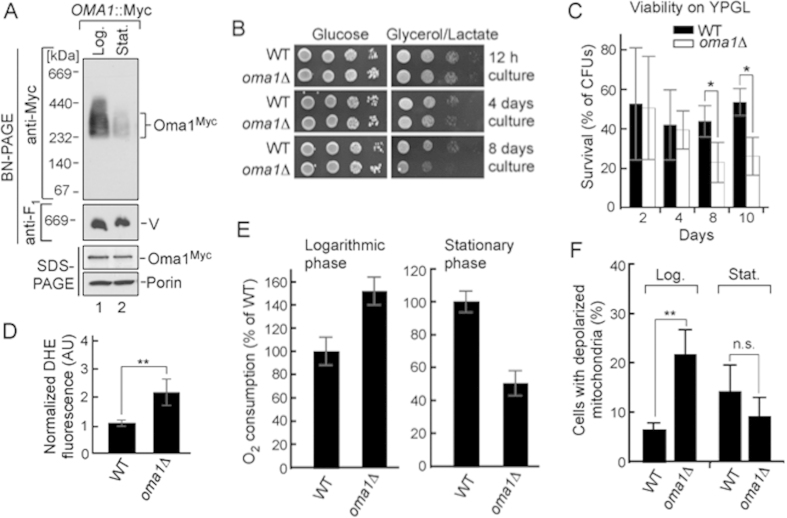
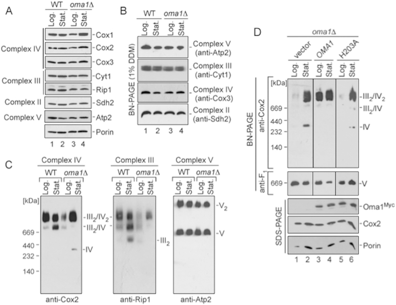
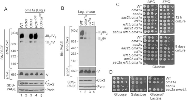
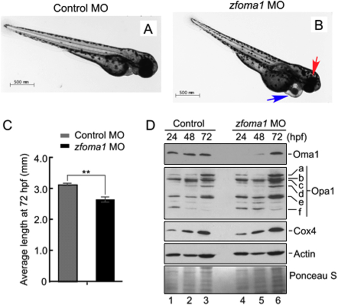
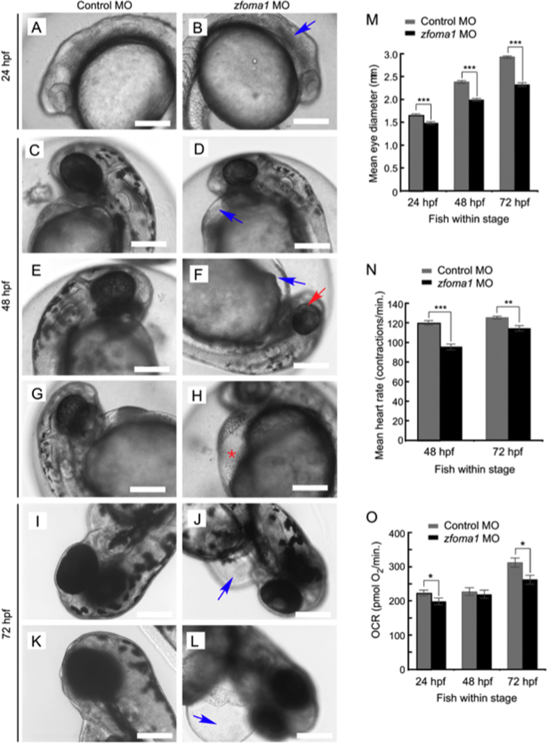
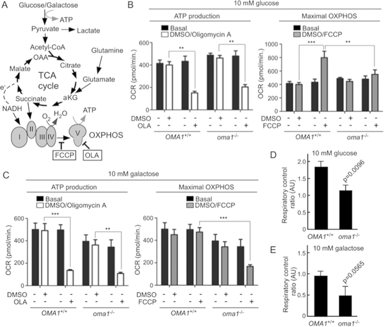
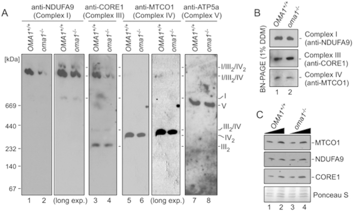
Mitochondria are involved in key cellular functions including energy production, metabolic homeostasis, and apoptosis. Normal mitochondrial function is preserved by several interrelated mechanisms. One mechanism - intramitochondrial quality control (IMQC) - is represented by conserved proteases distributed across mitochondrial compartments. Many aspects and physiological roles of IMQC components remain unclear. Here, we show that the IMQC protease Oma1 is required for the stability of the respiratory supercomplexes and thus balanced and tunable bioenergetic function. Loss of Oma1 activity leads to a specific destabilization of respiratory supercomplexes and consequently to unbalanced respiration and progressive respiratory decline in yeast. Similarly, experiments in cultured Oma1-deficient mouse embryonic fibroblasts link together impeded supercomplex stability and inability to maintain proper respiration under conditions that require maximal bioenergetic output. Finally, transient knockdown of OMA1 in zebrafish leads to impeded bioenergetics and morphological defects of the heart and eyes. Together, our biochemical and genetic studies in yeast, zebrafish and mammalian cells identify a novel and conserved physiological role for Oma1 protease in fine-tuning of respiratory function. We suggest that this unexpected physiological role is important for cellular bioenergetic plasticity and may contribute to Oma1-associated disease phenotypes in humans.


Figures

Figure 1
Figure 1. Deletion of Oma1 leads to progressive mitochondrial dysfunction.
(A) Electrophoretic analyses of mitochondria from wild-type (WT) strain with Oma1-13xMyc chromosomal tag isolated at exponential (12 h post-inoculation, A600 of 0.8; Log.) and stationary (48 h post-inoculation, A600 of 8.0; Stat.) phases of growth. Mitochondria (70 μg) were solubilized with 1.5% digitonin and subjected to BN-PAGE; 20 μg of mitochondria were used for SDS-PAGE. Oma1 was detected by immunoblotting with anti-Myc. Monomeric form of Complex V (V; BN-PAGE loading control) visualized with anti-F1 serum; porin was SDS-PAGE loading control. Source data (full-length blots) are available online in Supplementary information. (B) Respiratory growth of WT and oma1Δ strains. Synchronized cells were cultured in YPD medium for 0.5 (12 hours), 4 and 8 days at 28 °C and spotted onto YPD (Glucose) or YPGL (Glycerol/Lactate) plates. Pictures taken after 2 (YPD) or 4 (YPGL) days of growth at 28 °C. (C) WT and oma1Δ strains were grown in YPD medium for indicated number of days, diluted to 600 cells and plated on YPGL plates. Following a 4-days incubation at 28 °C, number of colony forming units was determined (n = 3 biologically independent experiments). The values were normalized to number of colonies that each strain formed when cultured on glucose-supplemented plates. (D) Endogenous superoxide levels in WT and oma1Δ cells. Log-phase cells stained with O2.−-specific dye dihydroethidium (DHE) were analyzed by flow cytometry (n = 4). (E) Oxygen consumption of synchronized WT and oma1Δ cells at log (A600 of 0.8) and stationary (A600 of 8.0) stages of growth; n = 3 independent cultures per each strain. (F) Mitochondrial membrane potential of WT and oma1Δ strains during log and stationary growth, assessed by flow cytometry analysis of JC-1-stained cells (n = 3). Data represent mean values ± S.D. **p < 0.01, *p < 0.05, n.s. =  non-significant (t-test).
Figure 2
Figure 2. Respiratory supercomplexes are impaired in Oma1-deficient yeast cells.
(A) Steady-state levels of the Cox1-Cox3 subunits of CcO (Complex IV), Cyt1 and Rip1 subunits of bc1cytochrome c reductase (Complex III), Sdh2 subunit of succinate dehydrogenase (Complex II), and Atp2 subunit of F1FO ATP synthase (Complex V), and porin were assessed by immunobloting of mitochondria (20 μg) from WT and oma1Δ cells. (B) BN-PAGE of individual ETC complexes from log and stationary phase WT and oma1Δ cells. Mitochondria (40 μg) were solubilized with 1% dodecyl maltoside (DDM). The complexes were visualized by blotting with indicated antibodies. (C) BN-PAGE of the above mitochondria lysed with 1.5% digitonin. (D) oma1Δ cells bearing vector, Myc-tagged Oma1 or its H203A variant were grown in synethetic galactose medium and used for mitochondrial isolation. Mitochondria (70 μg) were analyzed by BN-PAGE as in C. Another 20 μg of mitochondria were used for SDS-PAGE. Source data (full-length blots) are available online in Supplementary information.
Figure 3
Figure 3. OMA1 genetically interacts with supercomplex-stabilizing factors.
(A) BN- and SDS-PAGE analysis of mitochondria from log-phase oma1Δ cells expressing OMA1, overexpressing RCF1 or COX5a. Source data (full-length blots) are available online in Supplementary information. (B) Mitochondria from log-phase wild-type (WT), oma1Δ rcf1Δ and rcf1Δ oma1Δ cells were analyzed by native (70 μg of mitochondria) or denaturing (20 μg of mitochondria) PAGE. (C) Indicated strains were cultured in YPD medium for 0.5 (12 hours) and 8 days at 28 °C, spotted onto YPD (Glucose) plates and incubated at 28 °C or 37 °C. Pictures were taken after 2 days of growth. (D) The indicated strains were dropped onto YPD (Glucose), YPGal (Galactose) and YPGL (Glycerol/Lactate) plates and incubated at 28 °C.
Figure 4
Figure 4. Depletion of Oma1 in fish affects development.
(A,B) Representative images of larval zebrafish at 72 hours post fertilization (hpf) injected with either a standard control MO (A) or a zfoma1 MO (B). Fish injected with zfoma1 MO have smaller heads and eyes (red arrow) and defects in heart morphology (blue arrow). Bar, 500 μm. (C) Body length of control and zfoma1 morphants at 72 hpf. Average standard length was plotted ± S.E.M. (n = 6–13, **p < 0.01 by t-test). (D) Immunoblot of protein extracts from injected fish (yolk removed) at 24, 48 and 72 hpf. Expression of Oma1, Opa1 isoforms (a–f) and Cox4 (mitochondrial abundance marker) was analyzed with respective antibodies. Actin and Ponceau S staining were loading controls. Source data (full-length blots) are available online in Supplementary information.
Figure 5
Figure 5. Depletion of Oma1 in fish results in specific developmental abnormalities.
(A–L) At 24 hpf, zfoma1 morphants (B) lack definition of brain structures (blue arrow) compared to control MO fish (A). At 48 hpf, several abnormalities are observed in Oma1 MO-injected embryos (D,F,H). The embryos had smaller heads and eyes and often exhibited pericardial edema (blue arrow, D,F) that sometimes resulted in visible erythrocyte accumulation in the yolk sinus area (red asterisk, H). Pigmentation in the eye was partially complete compared to controls (red arrow, F). At 72 hpf, pericardial edema was extensive (blue arrow, J,L) and hearts were largely unlooped. Scale bars, 200 μm. (M) Eye size in control and zfoma1 morphants at 24, 48 and 72 hpf (n = 9–12). (N) Heart contraction rates (measured as beats per minute) in control versus Oma1-depleted fish at 48 and 72 hpf (n = 30). (O) In vivo respiration in control and Oma1 MO-treated fish embryos at 24, 48 and 72 hpf (n=10). Data are shown as mean ± S.E.M.; *p < 0.05, **p < 0.01, ***p < 0.001 zfoma1 vs. control MO (t-test).
Figure 6
Figure 6. Impaired bioenergetic function in oma1−/− MEFs.
(A) Schematic of cellular energy-converting pathways and inhibitors used to profile MEFs’ bioenergetics. FCCP, carbonyl cyanide 4-(trifluoromethoxy)phenylhydrazone; OLA, oligomycin A. (B,C) Oxygen consumption rates (OCR) in wild type (OMA1+/+) and oma1−/− MEFs under basal, OLA- and FCCP-stimulated conditions. Cells were cultured in the media containing 10 mM glucose (B) or 10 mM galactose (C). Data are shown as mean ± S.E.M. (n = 3 biological replicates); *p < 0.05, **p < 0.01, ***p < 0.001 (unpaired t-test). (D,E) Respiratory control ratios (a ratio between FCCP-stimulated OCR and basal OCR) in OMA1+/+ and oma−/− cells cultured in 10 mM glucose (D) or 10 mM galactose (E). Error bars indicate S.D.; p values are relative to control.
Figure 7
Figure 7. Loss of OMA1 impairs mammalian RSCs.
(A) BN-PAGE of mitochondria from wild type (OMA1+/+) and oma1−/− MEFs. Mitochondria (80 μg) were solubilized with 2% digitonin. Protein complexes were visualized with antibodies to NDUFA9 (Complex I), CORE1 (Complex III), MTCO1 (Complex IV) and ATP5A (Complex V). (B) BN-PAGE of WT and oma1−/− mitochondrial lysates. Mitochondria (40 μg) were solubilized with 1% dodecyl maltoside (DDM). Individual ETC complexes were visualized by immunoblotting with indicated antibodies. (C) Steady-state levels of the indicated subunits of ETC complexes in WT and oma1−/− mitochondria. Ten and 15 μg of mitochondria were analyzed by SDS-PAGE. Source data (full-length blots) for key panels of this figure are available online in Supplementary information.

References

    1. Vafai S. B. & Mootha V. K. Mitochondrial disorders as windows into an ancient organelle. Nature 491, 374–383 (2012). - PubMed
    1. Figueira T. R. et al. Mitochondria as a source of reactive oxygen and nitrogen species: from molecular mechanisms to human health. Antioxid. Redox Signal. 18, 2029–2074 (2013). - PubMed
    1. Wallace D. C. A mitochondrial paradigm of metabolic and degenerative diseases, aging and cancer: a dawn for evolutionary medicine. Ann. Rev. Genet. 39, 359–407 (2005). - PMC - PubMed
    1. Baker B. M. & Haynes C. M. Mitochondrial protein quality control during biogenesis and aging. Trends Biochem. Sci. 36, 254–261 (2011). - PubMed
    1. Vogel F., Bornhovd C., Neupert W. & Reichert A. S. Dynamic subcompartmentalization of the mitochondrial inner membrane. J. Cell Biol. 175, 237–247 (2006). - PMC - PubMed

Publication types

MeSH terms