. 2016 Jan 15:257:55-63.
doi: 10.1016/j.jneumeth.2015.09.002. Epub 2015 Sep 10.

Custom fit 3D-printed brain holders for comparison of histology with MRI in marmosets

Affiliations

Custom fit 3D-printed brain holders for comparison of histology with MRI in marmosets

Joseph R Guy et al. J Neurosci Methods. .

Abstract

Background: MRI has the advantage of sampling large areas of tissue and locating areas of interest in 3D space in both living and ex vivo systems, whereas histology has the ability to examine thin slices of ex vivo tissue with high detail and specificity. Although both are valuable tools, it is currently difficult to make high-precision comparisons between MRI and histology due to large differences inherent to the techniques. A method combining the advantages would be an asset to understanding the pathological correlates of MRI.

New method: 3D-printed brain holders were used to maintain marmoset brains in the same orientation during acquisition of ex vivo MRI and pathologic cutting of the tissue.

Results: The results of maintaining this same orientation show that sub-millimeter, discrete neuropathological features in marmoset brain consistently share size, shape, and location between histology and ex vivo MRI, which facilitates comparison with serial imaging acquired in vivo.

Comparison with existing methods: Existing methods use computational approaches sensitive to data input in order to warp histologic images to match large-scale features on MRI, but the new method requires no warping of images, due to a preregistration accomplished in the technique, and is insensitive to data formatting and artifacts in both MRI and histology.

Conclusions: The simple method of using 3D-printed brain holders to match brain orientation during pathologic sectioning and MRI acquisition enables rapid and precise comparison of small features seen on MRI to their underlying histology.

Keywords: 3D printing; EAE; Ex vivo; Histology; MRI; Marmoset.


Figures

Figure 1
Figure 1
Generation of a marmoset brain 3D model. After a marmoset brain has been fixed in paraformaldehyde (A), a T2-weighted MRI is performed at a 150 μm isotropic resolution (B). The images are upscaled and thresholded to produce a binary volumetric image (C), the surface of which is used to generate a 3D model of the brain (D). Note: Brain pictured in B, C, and D (marmoset 1) is from a different animal than the brain pictured in A.
Figure 2
Figure 2
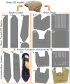
The brain model is used to build custom-fit brain holders. This figure shows the transition from the brain 3D model and blank objects to the final 3D models ready to print. The brain cradle (A) is based on the internal volume of a 50 mL Falcon tube. Views from the side (A1), top (A2), perspective (A3), and front (A4) are provided. The brain slicer is built to contain the brain and guide the blades during pathologic cutting. Perspective (B1), side (B2), front (B3), and top (B4) views are provided. The 3D marmoset brain model is centered on both the cradle (D1) and the slicer (C1,C2) before a Boolean subtraction is applied (D2,C3). Photographs of the brain cradle and slicer are shown with the brain in place in D3 and C4.
Figure 3
Figure 3
Brains from Marmoset 2 (A) and Marmoset 3 (B) are shown with their respective brain slicers. The blade holders with their blade inserts are shown in a configuration to cut all 2.5 mm thick brain slabs simultaneously (A) or to extract a single 5.0 mm thick slab of tissue containing a region of interest. Slicing downward and then retracting the blade-holder assembly extracted the tissue slab of interest (B).
Figure 4
Figure 4
Tissue slab photographs with corresponding MRI slices. Ex vivo MRI (column B) slices were predicted based on blade position in the 3D model and in vivo slices were visually matched (column A). After pathologic cutting of the tissue, photographs (column C) were found to be consistent with ex vivo MRI slices and comparable to in vivo MRI across different slabs of tissue in marmoset 1.
Figure 5
Figure 5
Whole slice matches between histology and ex vivo T2* MRI. Histology slices without significant artifact were selected approximately every 600 μm from each slab of tissue and displayed to the left of their corresponding MRI slice match. Two slabs from animal 1 (slab 1 A1-A4, slab 2 B1-B4), and one slab each from animal 2 (C1) and animal 3 (D1-D7) are shown. Whole-volume rotations of the MRI in the X and Y dimensions were necessary to increase precision: 0° for slab 1 animal 1, 3.6° for slab 2 animal 1, 0° for animal 2, 4.1° for animal 3. Close inspection reveals consistently matching small features (white matter lesions and small white matter tracts that change position from slice to slice) as well as overall anatomic matching.
Figure 6
Figure 6
Selected MRI and histology slices show the correspondence of matching in both whole-slice and magnified views of EAE lesions. Two different marmoset brains and slices from different areas within those brains (A,B) were selected to show the high-detail preservation between in vivo MRI (A1, B1), final ex vivo MRI (A2, B2) and histology (A3, B3). In the first animal (panel A), the magnified views show that ex vivo MRI (A4) can be correlated on the voxel level to histology (A5). The location and shape of demyelinated EAE lesions (hyperintense areas on MRI and absence of blue LFB stain and increased cell density on histology) in the optic tract are matching, as indicated by the arrows pointing at the lesion borders. In the second animal (panel B), similar features can be observed on in vivo MRI (B1) ex vivo MRI (B2) and histology (B3). In the magnified views, the same EAE lesion was visualized on in vivo MRI (B4) in addition to the ex vivo MRI (B5) and histology (B6). Note the lesser degree of precision of the in vivo MRI due to its lower image resolution. Note also the hypointense focal signal (green asterisk) within the lesion observed only on the T2* ex vivo MRI, which most likely represents an area of local iron (hemosiderin) deposition.
Figure 7
Figure 7
High-magnification views of corresponding MRI and histology. Magnified views were selected from images in Figure 5 to show that the high-precision matching between the H&E+LFB histology stain (A series) and the T2* ex vivo MRI (B series) is consistent throughout different areas of different brains. MRI images were windowed to best display lesion borders and histology images were rotated and resized to match. Arrows delineate lesions of high intensity on the MRI and their correspondence to absence of LFB staining (pink) and cellular infiltration (purple) on the histology images. Green asterisks indicate exceptionally low intensity regions within lesions that may represent local iron (hemosiderin) deposition.

References

    1. Boretius S, Schmelting B, Watanabe T, Merkler D, Tammer R, Czéh B, et al. Fuchs E. Monitoring of EAE onset and progression in the common marmoset monkey by sequential high-resolution 3D MRI. NMR in Biomedicine. 2006;19(1):41–49. - PubMed
    1. Gaitán MI, Maggi P, Wohler J, Leibovitch E, Sati P, Calandri IL, et al. Reich DS. Perivenular brain lesions in a primate multiple sclerosis model at 7-tesla magnetic resonance imaging. Multiple Sclerosis Journal. 2014;20(1):64–71. - PMC - PubMed
    1. Leibovitch E, Wohler JE, Macri SMC, Motanic K, Harberts E, Gaitán MI, et al. Jacobson S. Novel marmoset (Callithrix jacchus) model of human Herpesvirus 6A and 6B infections: immunologic, virologic and radiologic characterization. PLoS pathogens. 2013;9(1):e1003138. - PMC - PubMed
    1. Rausch M, Hiestand P, Baumann D, Cannet C, Rudin M. MRI-based monitoring of inflammation and tissue damage in acute and chronic relapsing EAE. Magnetic resonance in medicine. 2003;50(2):309–314. - PubMed
    1. ‘t Hart BA, Bauer J, Muller HJ, Melchers B, Nicolay K, Brok H, et al. Massacesi L. Histopathological characterization of magnetic resonance imaging-detectable brain white matter lesions in a primate model of multiple sclerosis: a correlative study in the experimental autoimmune encephalomyelitis model in common marmosets (Callithrix jacchus) The American journal of pathology. 1998;153(2):649–663. - PMC - PubMed

Publication types

MeSH terms