. 2015 Sep 17;59(6):931-40.
doi: 10.1016/j.molcel.2015.07.027. Epub 2015 Sep 10.

P16INK4a Upregulation Mediated by SIX6 Defines Retinal Ganglion Cell Pathogenesis in Glaucoma

Affiliations

P16INK4a Upregulation Mediated by SIX6 Defines Retinal Ganglion Cell Pathogenesis in Glaucoma

Dorota Skowronska-Krawczyk et al. Mol Cell. .

Abstract

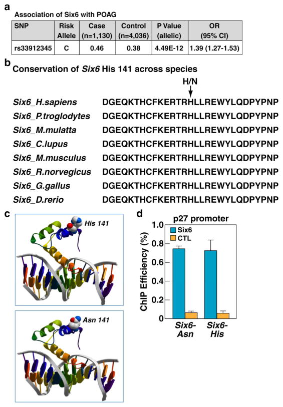
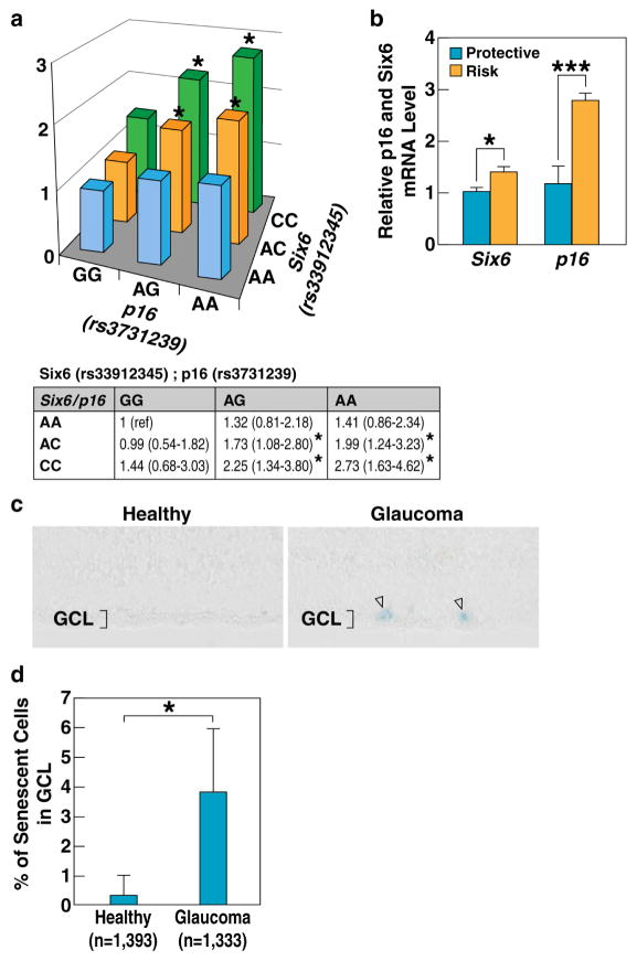
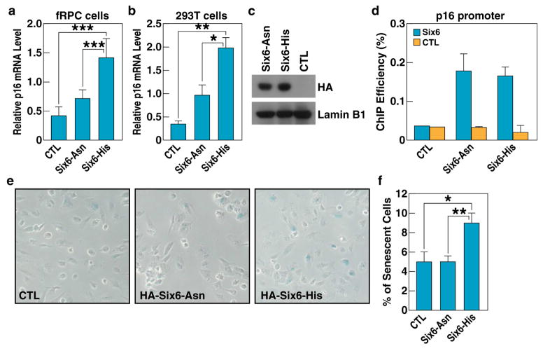
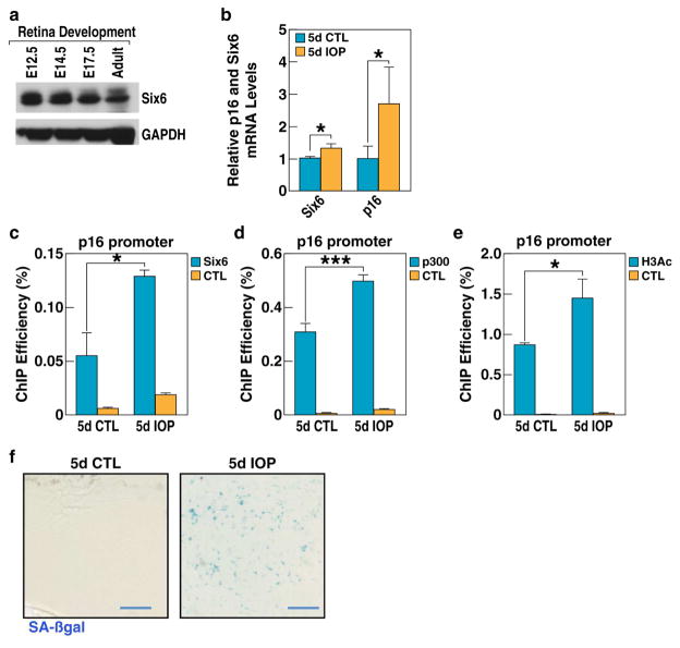
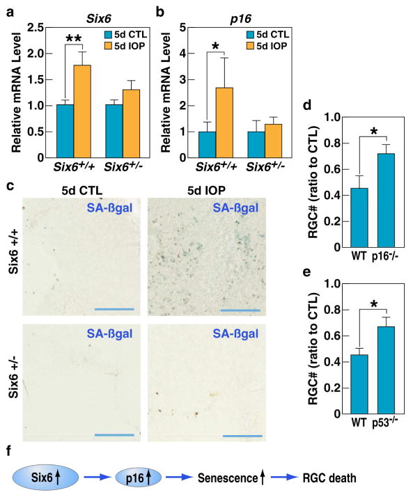
Glaucoma, a blinding neurodegenerative disease, whose risk factors include elevated intraocular pressure (IOP), age, and genetics, is characterized by accelerated and progressive retinal ganglion cell (RGC) death. Despite decades of research, the mechanism of RGC death in glaucoma is still unknown. Here, we demonstrate that the genetic effect of the SIX6 risk variant (rs33912345, His141Asn) is enhanced by another major POAG risk gene, p16INK4a (cyclin-dependent kinase inhibitor 2A, isoform INK4a). We further show that the upregulation of homozygous SIX6 risk alleles (CC) leads to an increase in p16INK4a expression, with subsequent cellular senescence, as evidenced in a mouse model of elevated IOP and in human POAG eyes. Our data indicate that SIX6 and/or IOP promotes POAG by directly increasing p16INK4a expression, leading to RGC senescence in adult human retinas. Our study provides important insights linking genetic susceptibility to the underlying mechanism of RGC death and provides a unified theory of glaucoma pathogenesis.


Figures

Figure 1
Figure 1. SIX6 protein residue 141 variants bind to DNA with similar efficiency
a, Association of SIX6 risk variant with POAG. Shown are the frequencies of SIX6 141 amino acid variants in Caucasian population in correlation with POAG. b, Conservation of SIX6 His141 (risk) variant across the species, Ans141 (protective) variant is present only in human lineage. c, Computer modeling of SIX6 structure. Upper panel: model of SIX6 with histidine at position 141; lower panel: model of SIX6 with asparagine at position 141. d, ChIP-qPCR analysis of SIX6 binding to p27 regulatory element in patient-derived lymphoblastoid cells shows similar efficiency of binding of both SIX6 variants. Experiments repeated 3 times, +/− SD. CTL, negative control; See also Figure S1.
Figure 2
Figure 2. Joint effect of specific alleles of SIX6 (rs33912345) and P16/INK4A (rs3731239) suggest functional interaction between the two genes
a, Results of the logistic regression analysis, plotted as Z-axis by odds ratios. b, RT-qPCR analysis of mRNA expression of SIX6 and P16/INK4A in human lymphocytes stratified by their SIX6 (rs33912345) genotypes. Four cell lines with rs33912345-AA (non-risk alleles) and four cell lines with rs33912345-CC (risk alleles) were analyzed. Relative mRNA levels were calculated by normalizing results with GAPDH and expressed relative to the AA genotype. p-values were calculated using two-tailed Student’s t-test. (+/− SD; *p<0.05, ***p<0.001). c, SA-βgal staining of human retinas indicating higher numbers of senescent cells in retinas with POAG. d, Quantification of senescent cells in healthy and POAG retinas (*p<0.05); See also Figure S2.
Figure 3
Figure 3. Increased expression of SIX6-risk variant correlates with a higher senescence rate
a, RT-qPCR analysis shows that overexpression of SIX6-His variant increased p16/INK4A expression in fRPCs. Experiments repeated 3 times, p-values were calculated using two-tailed Student’s t-test. (+/− SD, ***p<0.001). CTL, negative control b, RT-qPCR analysis shows that the overexpression of the SIX6-His variant increased P16/INK4A expression in 293T cells. Experiments repeated 3 times, p-values calculated using a two-tailed Student’s t-test. (+/− SD; *p<0.05, **p<0.01). CTL, negative control c, Western-blot confirming similar levels of expression of both SIX6 variants in transient transfections experiments. CTL, negative control d, ChIP-qPCR analysis of SIX6 variant association with P16/INK4A promoter, showing similar level of binding. CTL, negative control e, SA-βgal staining of the fRPCs transfected with either of the two SIX6 variants showing higher ratio of senescence in cells transfected with SIX6-141His risk variant. CTL, negative control f, Quantification of β-galactosidase-positive cells in fRPCs transfected with SIX6 variants. p-values calculated using a two-tailed Student’s t-test. (+/− SD; *p<0.05, **p<0.01). CTL, negative control; See also Figure S3.
Figure 4
Figure 4. Increased expression of SIX6 and induction of cell senescence in retinas upon IOP elevation
a, The expression of SIX6 protein in mouse retina during development and in the adult stage analyzed by Western blotting. b, RT-qPCR analysis of Six6 and P16/INK4A mRNA levels shows elevated expression of SIX6 and P16/INK4A in IOP-elevated mouse retinas 5 days after induction of acute experimental glaucoma (5d IOP) as compared to non-treated retina (5d CTL). Experiments were repeated in 8 animals, p-values calculated using a two-tailed Student’s t-test. (+/− SD; *p<0.05). c, ChIP-qPCR analysis of SIX6 protein binding shows its higher association with the P16/INK4A promoter in retinas subjected to acute intraocular pressure increase (5d IOP) as compared to non-treated retina (5d CTL). Experiments repeated 3 times, p-values calculated using a two-tailed Student’s t-test. (+/− SD; *p<0.05). CTL, negative control. d, ChIP-qPCR analysis shows higher levels of p300 association with P16/INK4A promoter upon experimental glaucoma (5d IOP) as compared to non-treated retina (5d CTL). Experiments repeated 3 times, p-values calculated using a two-tailed Student’s t-test. (+/− SD; ***p<0.001). CTL, negative control. e, ChIP-qPCR shows higher level of H3 acetylation at P16/INK4A promoter after acute intraocular pressure increase (5d IOP) as compared to non-treated retina (5d CTL). Experiments repeated 3 times, p-values calculated using a two-tailed Student’s t-test (+/− SD; *p<0.05). CTL, negative control. f, SA-βgal staining of flat mount retinas isolated from treated and non-treated eyes. High number of senescent cells is evident in IOP treated tissue. See also Figure S4.
Figure 5
Figure 5. IOP elevation primarily affects retinal ganglion cells
a, SA-βgal staining of cross-sections in IOP treated (5d IOP) or non-treated (5d CTL) retinas. Senescent cells are localized in the ganglion cell layer (GCL). b, Double staining of IOP treated (5d IOP) or non-treated (5d CTL) flat mount retinas with SA-βgal and BRN3a antibodies. Most senescent cells are also BRN3a positive. c, Immunostaining of IOP-treated Thy1-CFP retinas using anti-GFP antibody. Majority of SA-βgal positive cells are also Thy1-CFP positive. d, Schematic diagram of immunopanning, e, His but not the Asn version of Six6 significantly upregulates p16/INK4A expression as compared to non-transfected (RGC-CTL) or GFP-transfected (RGC-GFP) purified rat retinal ganglion cells. p-values calculated using a two-tailed Student’s t-test (+/− SD; *p<0.05). See also Figure S5.
Figure 6
Figure 6. Absence of either Six6 or P16 protects against RGC death in glaucoma
a, b, RT-qPCR analysis of Six6 (a) and P16/INK4A (b) mRNA levels upon IOP treatment (5d IOP) shows elevated expression of Six6 and P16/INK4A only in wild-type (Six6+/+) retinas and not in Six6+/− retinas as compared to non-treated (5d CTL) retinas. Experiments repeated in 8 animals, p-values calculated using a two-tailed Student’s t-test. (+/− SD; *p<0.05, **p<0.01). c, SA-β-galactosidase staining of flat mount retinas isolated from IOP-treated and non-treated eyes of Six6+/+ and Six6+/− mice shows a lack of senescent cells in the treated tissue isolated from heterozygous mice (blue bar). d, Quantification of the RGC ratio in treated and non-treated retinas in WT and P16 −/− mice. e, Quantification of the RGC ratio in IOP treated and non-treated retinas in WT and p53 KO mice. f, Model of the sequence of events leading to RGC death upon Six6 upregulation in glaucoma. See also Figure S6.

Comment in

  • Blindness: Assassins of eyesight.
    Huberman AD, El-Danaf RN. Huberman AD, et al. Nature. 2015 Nov 26;527(7579):456-7. doi: 10.1038/527456a. Nature. 2015. PMID: 26607542 No abstract available.

References

    1. Aldahmesh MA, Khan AO, Hijazi H, Alkuraya FS. Homozygous truncation of SIX6 causes complex microphthalmia in humans. Clinical genetics. 2013;84:198–199. - PubMed
    1. Anderson AM, Weasner BM, Weasner BP, Kumar JP. Dual transcriptional activities of SIX proteins define their roles in normal and ectopic eye development. Development. 2012;139:991–1000. - PMC - PubMed
    1. Baker DJ, Wijshake T, Tchkonia T, LeBrasseur NK, Childs BG, van de Sluis B, Kirkland JL, van Deursen JM. Clearance of p16Ink4a-positive senescent cells delays ageing-associated disorders. Nature. 2011;479:232–236. - PMC - PubMed
    1. Burdon KP, Macgregor S, Hewitt AW, Sharma S, Chidlow G, Mills RA, Danoy P, Casson R, Viswanathan AC, Liu JZ, et al. Genome-wide association study identifies susceptibility loci for open angle glaucoma at TMCO1 and CDKN2B-AS1. Nature genetics. 2011;43:574–578. - PubMed
    1. Burton DG. Cellular senescence, ageing and disease. Age (Dordr) 2009;31:1–9. - PMC - PubMed

Publication types

MeSH terms