. 2015 Oct 6;23(10):1934-1942.
doi: 10.1016/j.str.2015.08.006. Epub 2015 Sep 10.

Structural Basis of Telomerase Inhibition by the Highly Specific BIBR1532

Affiliations

Structural Basis of Telomerase Inhibition by the Highly Specific BIBR1532

Christopher Bryan et al. Structure. .

Abstract

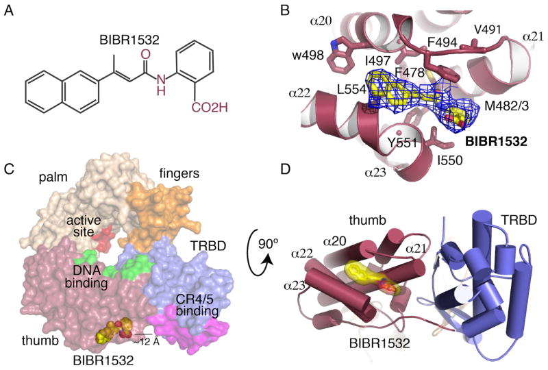
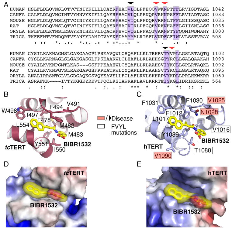
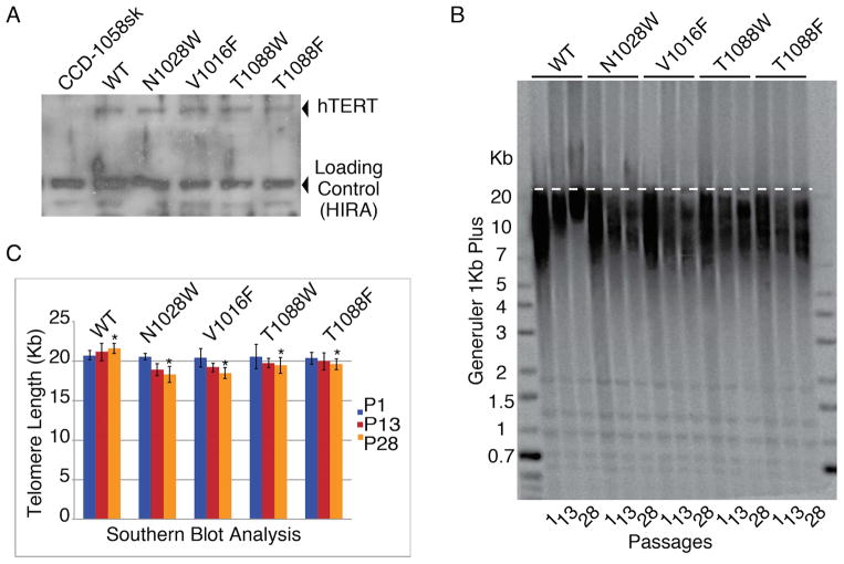
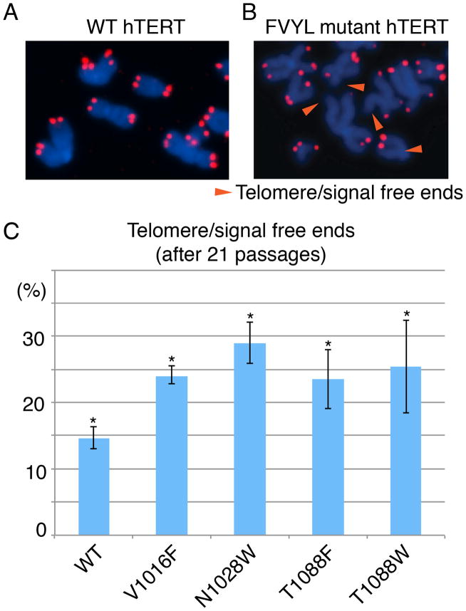
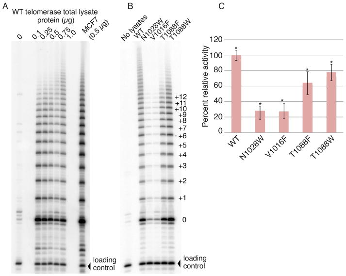
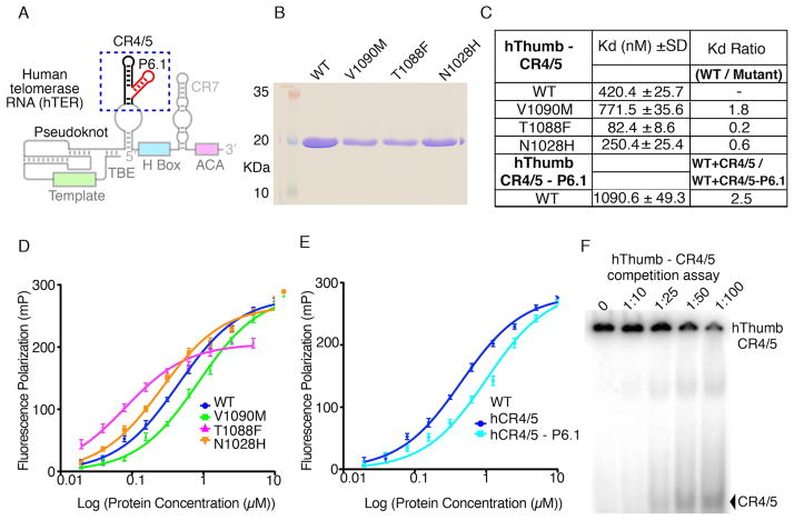
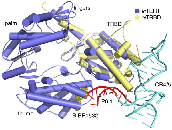
BIBR1532 is a highly specific telomerase inhibitor, although the molecular basis for inhibition is unknown. Here we present the crystal structure of BIBR1532 bound to Tribolium castaneum catalytic subunit of telomerase (tcTERT). BIBR1532 binds to a conserved hydrophobic pocket (FVYL motif) on the outer surface of the thumb domain. The FVYL motif is near TRBD residues that bind the activation domain (CR4/5) of hTER. RNA binding assays show that the human TERT (hTERT) thumb domain binds the P6.1 stem loop of CR4/5 in vitro. hTERT mutations of the FVYL pocket alter wild-type CR4/5 binding and cause telomere attrition in cells. Furthermore, the hTERT FVYL mutations V1025F, N1028H, and V1090M are implicated in dyskeratosis congenita and aplastic anemia, further supporting the biological and clinical relevance of this novel motif. We propose that CR4/5 contacts with the telomerase thumb domain contribute to telomerase ribonucleoprotein assembly and promote enzymatic activity.


Figures

Figure 1
Figure 1. Structure of the tcTERT - BIBR1532 complex
(A) Molecular structure of BIBR1532. (B) Simulated annealing (SA) omit map (blue mesh) of BBR1532 (yellow stick) bound to the tcTERT thumb domain; residues interacting with the inhibitor are shown in stick. (C) Surface representation of tcTERT showing the active site in red, the thumb DNA binding site in green and the TRBD CR4/5 binding site in magenta. BIBR1532 is shown in yellow spheres (D) Cartoon representation of tcTERT, panel C rotated 90° - BIBR1532 (yellow surface/stick) complex. Panels C and D show BIBR1532 binds in proximity of the TRBD-CR4/5 binding interface.
Figure 2
Figure 2. Structural comparison of the tcTERT and hTERT FVYL pockets
(A) Sequence alignment highlighting (purple color) the residues of tcTERT and hTERT involved in BIBR1532 binding. Residues mutated in this study are shown with an arrow. Naturally occurring missense mutations implicated in human disease are indicated with a red arrow. (B and C) Structural comparison of the tcTERT (crystal structure) and hTERT (PNAS model) FVYL pockets in complex with BIBR1532 (yellow stick); residues involved in inhibitor binding are shown in stick. Black boxes indicate hTERT residues mutated in cell-based and RNA binding assays. Residues associated with human disease are shown in red boxes. (D and E) Electrostatic surface representation of tcTERT and hTERT showing BIBR1532 bound to the FVYL pocket for comparison.
Figure 3
Figure 3. Southern blots analysis of CCD-1058Sk infected cells with wild type and FVYL, mutant hTERTs
(A) Western blot showing expression levels of WT and mutant hTERT proteins in CCD-1058Sk cells. (B) Southern blot analysis of FVYL pocket WT and mutant telomerases showing clear loss of telomere length with time (passages 1, 13, and 28). (C) Bar graph analysis of the telomere length for each celline at passages 1, 13 and 28 (TeloTool (MATLAB)) (Gohring et al, 2014). The standard deviation (error) associated with measurements and the statistical significance of the data are shown; *P<0.01 (two-tailed paired Student’s t-test).
Figure 4
Figure 4. Fluorescence in situ hybridization (FISH) data of CCD-1058Sk infected cells with wild type and FVYL, mutant hTERTs
FISH data at passage 21 showing chromosomes of CCD-1058Sk cells transformed with (A) WT hTERT and (B) FVYL pocket mutant hTERT; chromosomes with free telomere ends are indicated with an orange arrow; (C) Percentage of telomere/signal free ends in WT and FVYL mutant hTERT transfected CCD-1058Sk cells. The standard deviation (error) associated with an average of 1,500 chromosome measurements for each cell line carrying the WT or each of the FVYL pocket mutant hTERTs and the statistical significance of the data are shown; *P<0.05 (two-tailed paired Student’s t-test).
Figure 5
Figure 5. Telomerase activity assays of CCD-1058Sk infected cells with wild type and FVYL, mutant hTERTs
(A) TRAP activity of WT telomerase in 0, 0.25, 0.5, 0.75 and 1 μg total lysate protein concentration. (B) TRAP gel showing the activity of WT telomerase alongside the mutants N1028W, V1016F, T1088F, and T1088W in 0.5 μg of total lysate protein (C) Bar graph showing the relative activity, in terms of band intensity, of the wild type and FVYL mutant telomerases. The standard deviation (error) associated with 3 measurements and the statistical significance of the data are shown; **P<0.005 (two-tailed paired Student’s t-test).
Figure 6
Figure 6. Human thumb domain - RNA binding data
(A) Schematic of the hTER RNA showing conserved motifs in color. The CR4/5 and P6.1 motifs are highlighted with a blue dashed box. The template, pseudoknot and other conserved regions are also shown faded. (B) SDS PAGE gel of the WT and mutant hThumb proteins used in this study. (C) Table of WT and mutant hThumb-TBE binding constants calculated using Prism5 (GraphPad Software). (D) FP data of the CR4/5 with WT, N1028H, T1088F and V1090M hThumb proteins. (E) FP data of the CR4/5 minus P6.1 stem loop with WT hThumb. (F) SDS page gel of the competition assay of hThumb – CR4/5 complex with cold CR4/5.
Figure 7
Figure 7. Model of TERT, CR4/5 binding
Overlay of the crystal structures of tcTERT (PDB ID: 3DU6) and olTRBD-CR4/5 (PDB ID: 4026). tcTERT is shown in blue, the olTRBD in yellow and CR4/5 in cyan. The P6.1 stem loop of CR4/5 interacting with the FVYL pocket is shown in red.

References

    1. Banik SS, Guo C, Smith AC, Margolis SS, Richardson DA, Tirado CA, Counter CM. C-terminal regions of the human telomerase catalytic subunit essential for in vivo enzyme activity. Molecular and cellular biology. 2002;22:6234–6246. - PMC - PubMed
    1. Bley CJ, Qi X, Rand DP, Borges CR, Nelson RW, Chen JJ. RNA-protein binding interface in the telomerase ribonucleoprotein. Proceedings of the National Academy of Sciences of the United States of America. 2011;108:20333–20338. - PMC - PubMed
    1. Bradford MM. A rapid and sensitive method for the quantitation of microgram quantities of protein utilizing the principle of protein-dye binding. Analytical Biochemistry. 1976;72:248–254. - PubMed
    1. Damm K, Hemmann U, Garin-Chesa P, Hauel N, Kauffmann I, Priepke H, Niestroj C, Daiber C, Enenkel B, Guilliard B, Lauritsch I, Muller E, Pascolo E, Sauter G, Pantic M, Martens UM, Wenz C, Lingner J, Kraut N, Rettig WJ, Schnapp A. A highly selective telomerase inhibitor limiting human cancer cell proliferation. The EMBO journal. 2001;20:6958–6968. - PMC - PubMed
    1. El-Daly H, Kull M, Zimmermann S, Pantic M, Waller CF, Martens UM. Selective cytotoxicity and telomere damage in leukemia cells using the telomerase inhibitor BIBR1532. Blood. 2005;105:1742–1749. - PubMed

Publication types

MeSH terms

Associated data