. 2015 Sep 14:6:8327.
doi: 10.1038/ncomms9327.

Oxidation of the alarmin IL-33 regulates ST2-dependent inflammation

Affiliations

Oxidation of the alarmin IL-33 regulates ST2-dependent inflammation

E Suzanne Cohen et al. Nat Commun. .

Abstract

In response to infections and irritants, the respiratory epithelium releases the alarmin interleukin (IL)-33 to elicit a rapid immune response. However, little is known about the regulation of IL-33 following its release. Here we report that the biological activity of IL-33 at its receptor ST2 is rapidly terminated in the extracellular environment by the formation of two disulphide bridges, resulting in an extensive conformational change that disrupts the ST2 binding site. Both reduced (active) and disulphide bonded (inactive) forms of IL-33 can be detected in lung lavage samples from mice challenged with Alternaria extract and in sputum from patients with moderate-severe asthma. We propose that this mechanism for the rapid inactivation of secreted IL-33 constitutes a 'molecular clock' that limits the range and duration of ST2-dependent immunological responses to airway stimuli. Other IL-1 family members are also susceptible to cysteine oxidation changes that could regulate their activity and systemic exposure through a similar mechanism.


Conflict of interest statement

C.E.B. has received research support and consultancy fees from GSK, MedImmune, BI, Novartis, Roche, and Chiesi and has received travel support from BI, the European Respiratory Society, and the American Thoracic Society. The remaining authors are employees of the AstraZeneca Group and have stock/stock options in AstraZeneca.

Figures
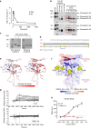
Figure 1
Figure 1. IL-33 is inactivated by disulphide bonding.
(a) Concentration of IL-33 (mean±s.e.m.) in bronchoalveolar lavage fluid (BALF) following intranasal Alternaria (ALT) challenge of BALB/c mice (n=3 per group), representative of 2 independent studies. (b) Western blot analysis of the same samples (pooled per group) under reducing and non-reducing conditions. Controls are as follows: cell lysate, lysate of HEK cells transfected with full length mouse IL-33; R&D, truncated mouse IL-33 (R&D systems); pep, truncated mouse IL-33 (Peprotech); PBS (30 min), BALF from vehicle (PBS) challenged mice at 30 min timepoint. (c) Non-reduced SDS-PAGE of IL-33112–270 either untreated (−) or post treatment with cell culture media (Iscoves Modified Dulbeccos Media) (+). Monomeric IL-33 was purified prior to analysis. (d) The human IL-33 linear sequence illustrated with the position of the cysteines and the proposed disulphide bridges formed after media treatment. Disulphide mapping was performed on purified monomeric protein. Numbering is based on the full length IL-33 sequence. (e) Hydrogen-exchange mass spectrometry (HX-MS) analysis of monomeric IL-33 and DSB IL-33. Comparison of fractional hydrogen exchange (for deuterium) in IL-33 (left panel) and DSB IL-33 (right panel). Data are mapped onto the published IL-33 structure in both cases for comparison purposes. Gaps in sequence coverage where no HX-MS data could be obtained are highlighted in slate blue. Side chains of cysteine residues are displayed as sticks. (f) Structural model displaying the difference in fractional hydrogen exchange between IL-33 and DSB IL-33 overlaid with the ST2 binding sites (red and magenta). Only increased hydrogen exchange was observed for DSB IL-33 versus IL-33, with regions exhibiting the greatest differences of hydrogen exchange depicted in dark blue. (g) ST2 binding of human IL-33 (upper panel) and purified DSB IL-33 (lower panel) measured by surface plasmon resonance. (h) Signalling in human umbilical vein endothelial cells (HUVEC) stimulated by human IL-33 and purified DSB IL-33. NFκB p65/RelA translocation was measured at 30 min post stimulation. Data points are mean±s.e.m. of duplicate determinations, representative of 3 independent experiments.
Figure 2
Figure 2. IL-33 converts rapidly to a disulphide bonded form in vivo.
Change in human IL-33 after exposure to serum or cell culture media measured using (a) ELISAs selective for reduced and disulphide bonded (DSB) IL-33. Data points are mean±s.e.m. of duplicate determinations, representative of two independent experiments. (b) Western blot of the same samples (pooled per group). (c,d) Profile of IL-33 forms (mean±s.e.m.) in bronchoalveolar lavage fluid (BALF) following Alternaria challenge of (c) Wild-type BALB/c (n=3 per group) or (d) Humanised IL-33 mice (n=3 per group). (e) Quantitation of IL-33 isoforms in human lung tissue, Mean±s.e.m. for n=6 donors. (f) Profile of IL-33 forms spontaneously released from human lung explants. Supernatants were pooled from 8 replicates per time point for a single donor and IL-33 levels measured (mean of duplicate ELISA replicates±s.e.m.). Data are representative of 2 individual donors. (g) Quantitation of human IL-33 forms in human sputum samples from severe asthmatics. Spontaneous or induced sputum was collected during stable asthma (n=37; black) or exacerbation (n=35; red) and IL-33 levels measured (mean of duplicate assay replicates). Solid symbols denote samples where both reduced and DSB IL-33 were detectable. Solid symbol shape corresponds to individual subjects. LLOD, lower limit of detection.
Figure 3
Figure 3. Free cysteines control the conformational switch in IL-33.
(a) SDS–PAGE of human N-terminal His-Avi tagged IL-33112–270 following overnight treatment as indicated. (b) NMR analysis with overlay of the 1H–15N HMQC spectra for 15N-labelled human IL-33 following 0 and 10 h incubation in cell culture media without air (left panel) or with air (right panel). Media was degassed before addition of IL-33. No air was added (left panel) or air was pumped through the sample for 5 min (right panel), representative of two separate experiments. (c) Signalling in human umbilical vein endothelial cells (HUVEC) stimulated by untreated or media treated human IL-33112–270 WT or complete Cys→Ser mutant. NFκB p65/RelA translocation was measured at 30 min post stimulation. Data points are mean±s.e.m. of duplicate determinations, representative of three independent experiments. (d) Stimulation of IL-6 production from human cord blood derived mast cells with human IL-33112–270 WT or Cys→Ser mutant. Data points are mean±s.e.m. of duplicate determinations, representative of two independent experiments. (e,f) Concentration of BALF IL-13 (e) or eosinophil counts (f) following 3 consecutive daily intranasal challenges of BALB/c mice with PBS (n=4 per group), human IL-33 WT or Cys→Ser mutant (n=6 per group). Endpoint was 24 h after final challenge. Statistical comparisons were made using one way ANOVA with Bonferroni multiple comparisons test (*P<0.05, **P<0.01, ***P<0.001). (g) NFκB p65/RelA translocation in human umbilical vein endothelial cells (HUVEC) stimulated by L-cystine treated human IL-33112–270 WT or Cys→Ser mutants. Data points are mean±s.e.m. of duplicate determinations, representative of two independent experiments.
Figure 4
Figure 4. Other IL-1 family members are susceptible to oxidation.
SDS–PAGE of mature IL-1 family members following overnight treatment (P, PBS+0.1% BSA; M, cell culture media+0.1% BSA). Non-reducing conditions (top panel); reducing conditions (lower panel).

References

    1. Schmitz J. et al.. IL-33, an interleukin-1-like cytokine that signals via the IL-1 receptor-related protein ST2 and induces T helper type 2-associated cytokines. Immunity 23, 479–490 (2005). - PubMed
    1. Moussion C., Ortega N. & Girard J. P. The IL-1-like cytokine IL-33 is constitutively expressed in the nucleus of endothelial cells and epithelial cells in vivo: a novel 'alarmin'? PLoS ONE 3, e3331 (2008). - PMC - PubMed
    1. Pichery M. et al.. Endogenous IL-33 is highly expressed in mouse epithelial barrier tissues, lymphoid organs, brain, embryos, and inflamed tissues: in situ analysis using a novel Il-33-LacZ gene trap reporter strain. J. Immunol. 188, 3488–3495 (2012). - PubMed
    1. Kouzaki H., Iijima K., Kobayashi T., O'Grady S. M. & Kita H. The danger signal, extracellular ATP, is a sensor for an airborne allergen and triggers IL-33 release and innate Th2-type responses. J. Immunol. 186, 4375–4387 (2011). - PMC - PubMed
    1. Kakkar R., Hei H., Dobner S. & Lee R. T. Interleukin 33 as a mechanically responsive cytokine secreted by living cells. J. Biol. Chem. 287, 6941–6948 (2012). - PMC - PubMed

Publication types