

Contrast-enhanced Ultrasound in Detecting Endoleaks with Failed Computed Tomography Angiography Diagnosis after Endovascular Abdominal Aortic Aneurysm Repair
- PMID: 26365968
- PMCID: PMC4725553
- DOI: 10.4103/0366-6999.164935
Contrast-enhanced Ultrasound in Detecting Endoleaks with Failed Computed Tomography Angiography Diagnosis after Endovascular Abdominal Aortic Aneurysm Repair
Abstract
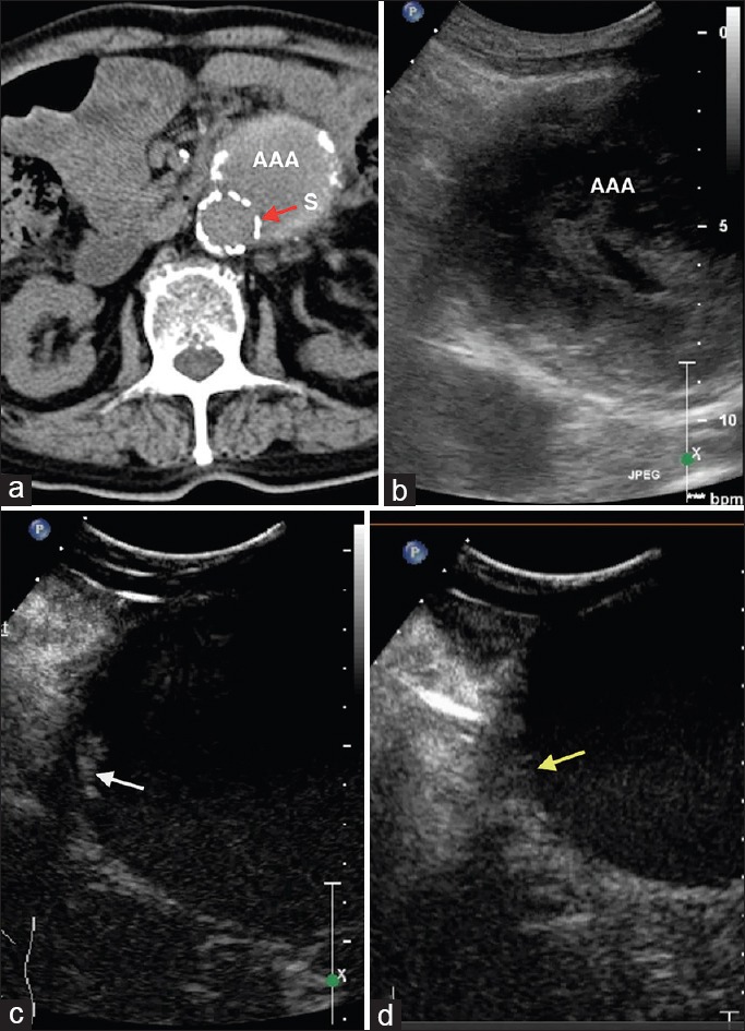
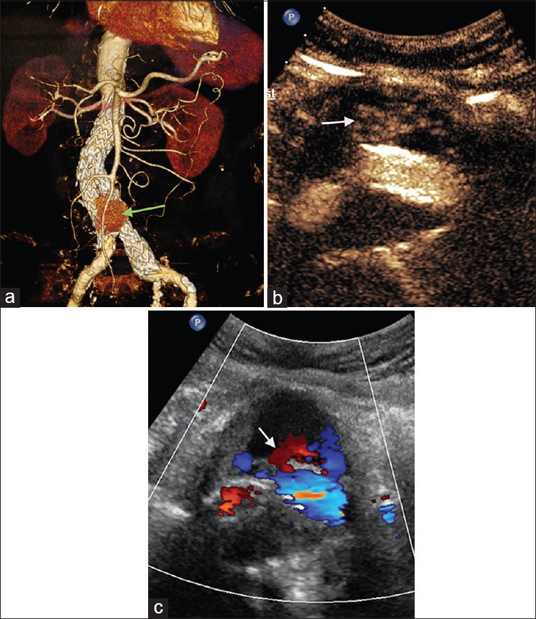
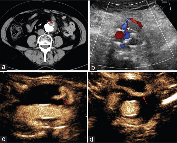
Background: Endovascular aneurysm repair (EVAR) is one of the first-line therapies of abdominal aortic aneurysms. Postoperative endoleak is the most common complication of EVAR. Computed tomography angiography (CTA), which is routine for follow-up, has side effects (e.g., radiation) and also has a certain percentage of missed diagnosis. Preliminary studies on contrast-enhanced ultrasound (CEUS) have shown that the sensitivity of CEUS for detecting endoleak is no lower than that of CTA. To investigate the advantages of CEUS, we conducted CEUS examinations of post-EVAR cases in which CTA failed to detect endoleak or could not verify the type of endoleak.
Methods: Post-EVAR patients, who were clinically considered to have endoleak and met the inclusion criteria were enrolled between March 2013 and November 2014. All of the patients underwent color Doppler flow imaging (CDFI) and a CEUS examination. Size, location, microbubble dispersion, and hemodynamic characteristics of leaks were recorded. Comparison between the diagnosis of CEUS and CDFI was conducted using Fisher's exact test and clinical outcomes of all patients were followed up.
Results: Sixteen patients were enrolled, and 12 (75%) had endoleaks with verified types by CEUS. Among 12 cases of endoleaks were positive by CEUS, 10 were CDFI-positive, and the four CEUS-negative cases were all negative by CDFI. The diagnostic values of CEUS and CDFI were statistically different (P = 0.008). Six patients with high-pressure endoleaks received endovascular re-intervention guided by CEUS results. One patient with type III endoleak had open surgery when endovascular repair failed.
Conclusions: CEUS is a new, safe, and effective means for detection of endoleaks post-EVAR. This technique can be used as a supplement for routine CTA follow-up to provide more detailed information on endoleak and its category.
Figures
References
-
- Nordon IM, Hinchliffe RJ, Loftus IM, Thompson MM. Pathophysiology and epidemiology of abdominal aortic aneurysms. Nat Rev Cardiol. 2011;8:92–102. - PubMed
-
- Moll FL, Powell JT, Fraedrich G, Verzini F, Haulon S, Waltham M, et al. Management of abdominal aortic aneurysms clinical practice guidelines of the European society for vascular surgery. Eur J Vasc Endovasc Surg. 2011;41(Suppl 1):S1–58. - PubMed
-
- Lederle FA, Freischlag JA, Kyriakides TC, Matsumura JS, Padberg FT, Jr, Kohler TR, et al. Long-term comparison of endovascular and open repair of abdominal aortic aneurysm. N Engl J Med. 2012;367:1988–97. - PubMed
-
- Mehta M, Byrne J, Darling RC, 3rd, Paty PS, Roddy SP, Kreienberg PB, et al. Endovascular repair of ruptured infrarenal abdominal aortic aneurysm is associated with lower 30-day mortality and better 5-year survival rates than open surgical repair. J Vasc Surg. 2013;57:368–75. - PubMed
Publication types
MeSH terms
Substances
LinkOut - more resources
Full Text Sources
Other Literature Sources
Miscellaneous
