Review
. 2015 Sep 2:10:5543-59.
doi: 10.2147/IJN.S83557. eCollection 2015.

Pre- and postmortem imaging of transplanted cells

Affiliations
Review

Pre- and postmortem imaging of transplanted cells

Anna Andrzejewska et al. Int J Nanomedicine. .

Abstract

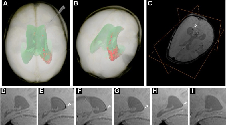
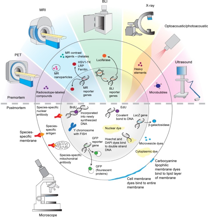
Therapeutic interventions based on the transplantation of stem and progenitor cells have garnered increasing interest. This interest is fueled by successful preclinical studies for indications in many diseases, including the cardiovascular, central nervous, and musculoskeletal system. Further progress in this field is contingent upon access to techniques that facilitate an unambiguous identification and characterization of grafted cells. Such methods are invaluable for optimization of cell delivery, improvement of cell survival, and assessment of the functional integration of grafted cells. Following is a focused overview of the currently available cell detection and tracking methodologies that covers the entire spectrum from pre- to postmortem cell identification.

Keywords: MRI; SPECT; bioluminescence; cell labeling; stem cells; transplantation.


Figures

Figure 1
Figure 1
Imaging of SPIO-labeled autologous cord blood derived cells in a patient with global cerebral ischemia. Notes: (A) Volume rendering of MRI data of the patient’s head obtained 24 hours posttransplantation. Semiautomatic segmentation is based on pixel intensity, showing the projection of the ventricular system (green) and the distribution of the SPIO signal from the transplanted cells within the occipital horn of the right ventricle (red). Note the supine configuration of the head, corresponding to positioning during surgery. The route and trajectory of cell transplantation via the frontal horn is represented by the needle. (B) Posterior-superior view of the patient’s head, emphasizing the location of the hypointense SPIO signal from autologous cord blood-derived cells transplanted within the occipital horn. (C) T2*-weighted image with an orthogonal view centered on the cellular SPIO signal in the occipital horn (white arrowhead). (DI) Sagittal T2*-weighted MRI scans showing a longitudinal dispersion of SPIO signal within the occipital horn (white arrowheads); (D) pretransplantation, (E) 24 hours posttransplantation (PT), (F) 7 days PT, (G) 2 months PT, (H) 4 months PT, and (I) 33 months PT. Abbreviations: SPIO, superparamagnetic iron nanoparticle; MRI, magnetic resonance imaging.
Figure 2
Figure 2
High-resolution MRI images. Notes: Brain has two tumors, a control (wt) and a glioma expressing a recombinant MRI reporter HSV1-TK, highlighted using CEST imaging. Abbreviations: MRI, magnetic resonance imaging; HSV1-TK, herpes simplex virus type-1 thymidine kinase; CEST, chemical exchange saturation transfer; h, hour.
Figure 3
Figure 3
Schematic outline of bioluminescence cells creation and their identification by BLI method in host body after transplantation. Abbreviation: BLI, bioluminescent imaging.
Figure 4
Figure 4
Depiction of the mechanism of optoacoustic imaging. Note: Copyright © 2015. Reproduced with permission from iThera Medical, (http://www.ithera-medical.com/technology/msot-principle.html).
Figure 5
Figure 5
The summary of pre- and postmortem labeling and identification techniques described in detail in text. Note: Reproduced with permission from © I-Hsun Wu 2014. Abbreviations: PET, positron emission tomography; BLI, bioluminescent imaging; MRI, magnetic resonance imaging; HSV1-TK, herpes simplex virus type-1 thymidine kinase; LRP, lysine-rich protein; MR, magnetic resonance; EdU, 5-ethynyl-2-deoxyuridine; FISH, fluorescence in situ hybridization; GFP, green fluorescent protein; DAPI, 4′6-diamidino-2-phenylindole; BrdU, 5-bromo-2′-deoxyuridine.

References

    1. Bhattacharyya S, Dixit M. Metallic radionuclides in the development of diagnostic and therapeutic radiopharmaceuticals. Dalton Trans. 2011;40(23):6112–6128. - PMC - PubMed
    1. Zeglis MB, Houghton JL, Evans MJ, Viola-Villegas N, Lewis JS. Underscoring the influence of inorganic chemistry on nuclear imaging with radiometals. Inorg Chem. 2014;53(4):1880–1899. - PMC - PubMed
    1. Mathiasen AB, Kastrup J. Non-invasive in-vivo imaging of stem cells after transplantation in cardiovascular tissue. Theranostics. 2013;3(8):561–572. - PMC - PubMed
    1. Kim MH, Wooa SK, Lee KC, et al. Longitudinal monitoring adipose- derived stem cell survival by PET imaging hexadecyl-4-124I-iodobenzoate in rat myocardial infarction model. Biochem Biophys Res Commun. 2015;456(1):13–19. - PubMed
    1. Janowski M, Bulte JW, Walczak P. Personalized nanomedicine advancements for stem cell tracking. Adv Drug Deliv Rev. 2012;64(13):1488–1507. - PMC - PubMed

Publication types