

Can a dual-energy computed tomography predict unsuitable stone components for extracorporeal shock wave lithotripsy?
- PMID: 26366277
- PMCID: PMC4565899
- DOI: 10.4111/kju.2015.56.9.644
Can a dual-energy computed tomography predict unsuitable stone components for extracorporeal shock wave lithotripsy?
Abstract
Purpose: To assess the potential of dual-energy computed tomography (DECT) to identify urinary stone components, particularly uric acid and calcium oxalate monohydrate, which are unsuitable for extracorporeal shock wave lithotripsy (ESWL).
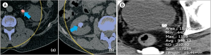
Materials and methods: This clinical study included 246 patients who underwent removal of urinary stones and an analysis of stone components between November 2009 and August 2013. All patients received preoperative DECT using two energy values (80 kVp and 140 kVp). Hounsfield units (HU) were measured and matched to the stone component.
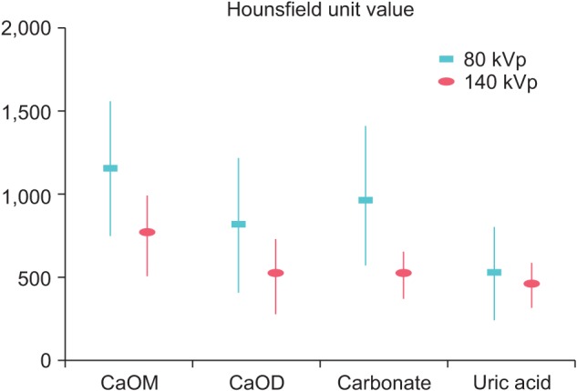
Results: Significant differences in HU values were observed between uric acid and nonuric acid stones at the 80 and 140 kVp energy values (p<0.001). All uric acid stones were red on color-coded DECT images, whereas 96.3% of the nonuric acid stones were blue. Patients with calcium oxalate stones were divided into two groups according to the amount of monohydrate (calcium oxalate monohydrate group: monohydrate≥90%, calcium oxalate dihydrate group: monohydrate<90%). Significant differences in HU values were detected between the two groups at both energy values (p<0.001).
Conclusions: DECT improved the characterization of urinary stone components and was a useful method for identifying uric acid and calcium oxalate monohydrate stones, which are unsuitable for ESWL.
Keywords: Calcium oxalate; Uric acid; Urinary calculi; X-ray computed tomography.
Conflict of interest statement
Figures
References
-
- Sourtzis S, Thibeau JF, Damry N, Raslan A, Vandendris M, Bellemans M. Radiologic investigation of renal colic: unenhanced helical CT compared with excretory urography. AJR Am J Roentgenol. 1999;172:1491–1494. - PubMed
-
- Miller OF, Rineer SK, Reichard SR, Buckley RG, Donovan MS, Graham IR, et al. Prospective comparison of unenhanced spiral computed tomography and intravenous urogram in the evaluation of acute flank pain. Urology. 1998;52:982–987. - PubMed
-
- Yilmaz S, Sindel T, Arslan G, Ozkaynak C, Karaali K, Kabaalioglu A, et al. Renal colic: comparison of spiral CT, US and IVU in the detection of ureteral calculi. Eur Radiol. 1998;8:212–217. - PubMed
-
- Niall O, Russell J, MacGregor R, Duncan H, Mullins J. A comparison of noncontrast computerized tomography with excretory urography in the assessment of acute flank pain. J Urol. 1999;161:534–537. - PubMed
-
- Wang JH, Shen SH, Huang SS, Chang CY. Prospective comparison of unenhanced spiral computed tomography and intravenous urography in the evaluation of acute renal colic. J Chin Med Assoc. 2008;71:30–36. - PubMed
MeSH terms
Substances
LinkOut - more resources
Full Text Sources
Other Literature Sources
Medical
