. 2015:1:15009.
doi: 10.1038/npjbiofilms.2015.9.

A unique biofilm in human deep mycoses: fungal amyloid is bound by host serum amyloid P component

Affiliations

A unique biofilm in human deep mycoses: fungal amyloid is bound by host serum amyloid P component

Melissa C Garcia-Sherman et al. NPJ Biofilms Microbiomes. 2015.

Abstract

Background/objectives: We have demonstrated the presence of Candida cell surface amyloids that are important in aggregation of fungi and adherence to tissue. Fungal amyloid was present in invasive human candidal infections and host serum amyloid P component (SAP) bound to the fungal amyloid. SAP is a protease-resistant glycoprotein that binds avidly to amyloid and interferes with host defence, especially against bacterial pathogens for which neutrophils are important. In this study, we investigated whether biofilm of fungal amyloid and SAP was a feature of other disseminated fungal infections.

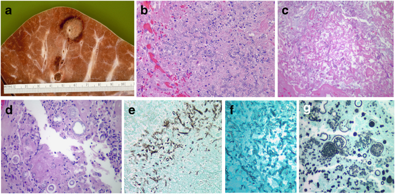
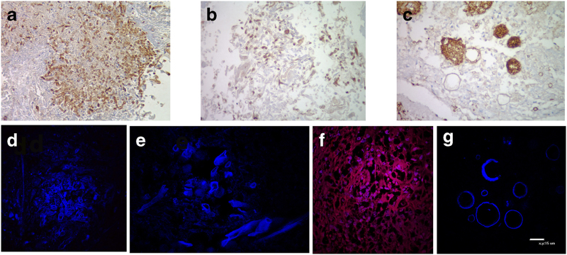
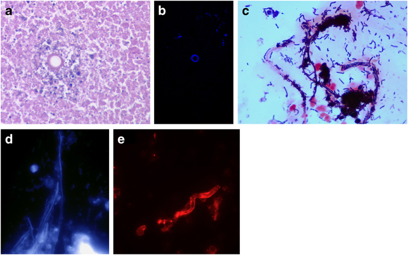
Methods: Tissue specimens from 15 autopsies were systematically evaluated with multiple histochemical stains including thioflavin T and Congo red (dyes that stain amyloid), as well as antibody to SAP. We studied specimens with disseminated aspergillosis, mucormycosis and coccidioidomycosis. The structure of the lesions, host inflammatory cells and the presence of fungal amyloid and SAP were determined.

Results: The structure of the lesions was characteristic in aspergillosis ('starburst') and mucormycosis (closely apposed bundles of hyphae). Host inflammatory cells were absent or few in number within these lesions. In Coccidioides lesions, host inflammation was sparse as well. Fungal amyloid was a prominent feature of all lesions along with abundant SAP bound to hyphae and spherules. Fungal amyloid and SAP perhaps contributed to persistence in caseous necrosis lesions. SAP also bound to Aspergillus and Mucorales amyloid in vitro.

Conclusions: A biofilm including amyloid and SAP is present in invasive fungal infections. This biofilm may dampen host defence leading to the characteristic sparse inflammatory reaction found in these infections.


Conflict of interest statement

The authors declare no conflict of interest.

Figures

Figure 1
Figure 1
Histology of fungal aggregates in situ. (a) View at autopsy of a stereotypical ‘starburst’ lesion of invasive Aspergillus of the lung (tan tissue) encircled by a halo of haemorrhage. (b) H&E stain demonstrating Aspergillus hyphae (light-blue structures in the centre of the photomicrograph) radiating through lung tissue and surrounded by a collar of haemorrhage. Note the absence of inflammatory cells around and within the tangle of fungal hyphae (×400). Compare b with e below it where darkly stained hyphae stand out from the acellular background. Hyphae are ~7 μ in diameter. (c) H&E stain of heart tissue with a myriad of ribbon-like, non-septate hyphae of a diameter of ~10 μ occupying the majority of the photomicrograph. These features are diagnostic of Mucorales species. Note the absence of host inflammatory cells within and surrounding the hyphal bundles. (d) H&E stain of lung tissue of a patient with pulmonary coccidioidomycosis demonstrating various sizes of spherules (20–50 μ) with and without endospores surrounded by a sparse inflammatory infiltrate (×400). (e) GMS of lung tissue showing Aspergillus hyphae (black structures) radiating outwards from a central focus or ‘starburst’ (×400). (f) GMS stain of heart showing large, black, non-septate hyphae. Note the density of the hyphae in the lesion. (g) GMS stain of lung tissue showing numerous spherules (black circles) and endospores (black dots); some spherules are empty and do not contain endospores, others are broken open and ‘spilling’ endospores (×400).
Figure 2
Figure 2
SAP and thioflavin T binding to fungal aggregates in situ. (a) Lung tissue stained with antibody to SAP that is bound to the wall of the Aspergillus hyphae (SAP stains a reddish-brown). This illustrates the spherical ‘starburst’ nature of the lesion (×400). (b) Heart tissue stained with antibody to SAP demonstrating patchy staining, often at junctions of the hyphae of Mucorales (×400). (c) Lung tissue stained with antibody to SAP showing heavy staining (reddish-brown) of endospores and spherule walls of Coccidioides (×400). (d) Thioflavin T stain of lung tissue containing hyphal elements of Aspergillus that are fluorescent and arranged in a ‘starbust;’ some hyphae are cut in cross-section and appear as tubular forms. The fluorescent structure within the hyphae is amyloid. (e) Thioflavin T stain of brain tissue demonstrating ribbon-like hyphae of Murcorales in oblique and cross-section. (f) Lung tissue stained with Congo red and thioflavin T demonstrating fluorescent endospores (numerous blue–red structures) originating from a spherule and scattered throughout lung tissue (stained red with Congo red). (g) Splenic tissue from a patient with disseminated coccidioidomycosis infiltrated with spherules that fluoresce with thioflavin T, demonstrating the amyloid in the spherule walls (×400).
Figure 3
Figure 3
SAP binding in situ and in vitro. (a) Lung tissue with a Coccidioides spherule in a granuloma undergoing caseous necrosis. The spherule is surrounded by eosinophilic cellular debris (H&E, ×400). (b) Same spherule stained with thioflavin T demonstrates the amyloid present in this empty, likely non-viable spherule that is impervious to degradation by the caseous process. (c) Gram stain of scraping from oropharyngeal thrush showing hyphae, yeast cells and Gram-positive bacteria adherent to and encasing the fungi with a paucity of host cells. (d) Thioflavin T stain of scraping from the same patient demonstrating amyloid in hyphae or pseudohyphae of Candida. (e) Condidia of Aspergillus flavus were cultured for 8 h in yeast extract peptone dextrose, washed and incubated in human serum for 2 h, followed by washing and staining for SAP with fluorescent antibody (red). Note: SAP decorated the entire hyphal surface.

Similar articles

Cited by

References

    1. Berenguer J , Allende MC , Lee JW , Garrett K , Lyman C , Ali NM et al. Pathogenesis of pulmonary aspergillus. Granulocytopenia versus cyclosporine and methylprednisolone-induced immunosuppression. Am J Resp Crit Care Med 1995; 152: 1079–1086. - PubMed
    1. Stergiopoulou T , Meletiadis J , Rollides E , Kleiner DE , Schaufele R , Roden M et al. Host-dependent patterns of tissue injury in invasive pulmonary aspergillosis. Am J Clin Pathol 2007; 127: 349–355. - PubMed
    1. Ben-Ami R , Luna M , Lewis R , Walsh T , Kontoyiannis D . A clinicopathological study of pulmonary mucormycosis in cancer patients: extensive angioinvasion but limited inflammatory response. J Infection 2009; 59: 134–138. - PMC - PubMed
    1. Gilchrist KB , Garcia MC , Sobonya RE , Lipke PN , Klotz SA . New features of invasive candidiasis in humans: amyloid formation by the fungi and deposition of serum amyloid P component by the host. J Infect Dis 2112; 206: 1473–1478. - PMC - PubMed
    1. Tennent GA , Lovat L , Pepys MB . Serum amyloid P component prevents proteolysis of amyloid fibers. Proc Natl Acad Sci USA 1995; 92: 4299–4303. - PMC - PubMed