Granulocyte-colony stimulating factor producing anaplastic carcinoma of the pancreas treated by distal pancreatectomy and chemotherapy: report of a case
- PMID: 26366343
- PMCID: PMC4560138
- DOI: 10.1186/s40792-015-0048-y
Granulocyte-colony stimulating factor producing anaplastic carcinoma of the pancreas treated by distal pancreatectomy and chemotherapy: report of a case
Abstract
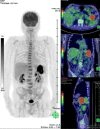
Granulocyte-colony stimulating factor (G-CSF) producing pancreatic cancers are extremely rare. These tumors have an aggressive clinical course but no established treatment. We encountered a patient with a G-CSF-induced pancreatic cancer who was treated by surgical resection, followed by steroid treatment and chemotherapy. A 68-year-old Asian male presented at a local hospital with a 3-month history of fever, loss of appetite, and 10-kg weight loss. Laboratory data showed leukocytosis and elevation of C-reactive protein. Computed tomography (CT) revealed a 50-mm mass in the tail of the pancreas, but no signs of infective foci. He was transferred to our hospital for further evaluation. Contrast-enhanced CT showed rapid growth of this tumor over 1 week, and (18) F-2-fluoro-2-deoxyglucose positron-emission tomography/computed tomography (FDG PET/CT) showed FDG accumulation in the tail of the pancreas (SUV max, 17.1) but at no other sites in his body. Magnetic resonance imaging showed a heterogeneous mass, similar to that observed by CT. Three weeks later, the patient underwent a distal pancreatectomy with splenectomy. The resected specimen was 154 mm in diameter, a threefold increase from the initial image. Histopathological examination identified the tumor as an anaplastic carcinoma of the pancreas. Following surgery, his leukocyte count and body temperature were reduced. He recovered well and was discharged from our hospital on postoperative day 18. Immunohistochemical expression of G-CSF in the resected specimen and elevated serum G-CSF concentration confirmed that the mass was a G-CSF producing anaplastic carcinoma of the pancreas. Subsequently, the patient experienced a high fever and loss of appetite. CT showed recurrence of cancer in the abdominal cavity, for which he was started immediately on tegafur-gimeracil-oteracil potassium combination S-1 and steroid. Unfortunately, he died on postoperative day 83. To our knowledge, this patient was the first with a G-CSF producing anaplastic carcinoma of the pancreas to be treated by surgical resection, steroid and adjuvant chemotherapy.
Keywords: Granulocyte-colony stimulating factor; Leukocytosis; Pancreatectomy; Pancreatic cancer; Steroid; TS-1.
Figures


Similar articles
-
A case of anaplastic carcinoma of the pancreas producing granulocyte-colony stimulating factor.Clin J Gastroenterol. 2009 Apr;2(2):109-114. doi: 10.1007/s12328-008-0058-4. Epub 2009 Jan 15. Clin J Gastroenterol. 2009. PMID: 26192175
-
Granulocyte Colony-stimulating Factor Producing Anaplastic Carcinoma of the Pancreas: Case Report and Review of the Literature.Anticancer Res. 2017 Jan;37(1):223-228. doi: 10.21873/anticanres.11310. Anticancer Res. 2017. PMID: 28011495
-
A case of anaplastic ductal carcinoma of the pancreas with production of granulocyte-colony stimulating factor.Hepatogastroenterology. 2006 Nov-Dec;53(72):957-9. Hepatogastroenterology. 2006. PMID: 17153462
-
[Resection of a Granulocyte Colony-Stimulating Factor-Producing Anaplastic Carcinoma of the Pancreas, Associated with Humoral Hypercalcemia of Malignancy].Gan To Kagaku Ryoho. 2018 May;45(5):859-862. Gan To Kagaku Ryoho. 2018. PMID: 30026452 Review. Japanese.
-
[Autopsy of anaplastic carcinoma of the pancreas producing granulocyte colony-stimulating factor].Nihon Shokakibyo Gakkai Zasshi. 2016 Aug;113(8):1408-15. doi: 10.11405/nisshoshi.113.1408. Nihon Shokakibyo Gakkai Zasshi. 2016. PMID: 27498938 Review. Japanese.
Cited by
-
Successful resection of a granulocyte colony-stimulating factor-producing carcinoma of the pancreas: A case report.Mol Clin Oncol. 2019 Oct;11(4):359-363. doi: 10.3892/mco.2019.1902. Epub 2019 Jul 22. Mol Clin Oncol. 2019. PMID: 31475063 Free PMC article.
-
Aggressive Recurrent Pancreatic Cancer Producing Granulocyte Colony-Stimulating Factor.Case Rep Gastroenterol. 2020 Jul 13;14(2):329-337. doi: 10.1159/000508439. eCollection 2020 May-Aug. Case Rep Gastroenterol. 2020. PMID: 32884507 Free PMC article.
-
Tumor-derived G-CSF induces an immunosuppressive microenvironment in an osteosarcoma model, reducing response to CAR.GD2 T-cells.J Hematol Oncol. 2024 Dec 18;17(1):127. doi: 10.1186/s13045-024-01641-7. J Hematol Oncol. 2024. PMID: 39695851 Free PMC article.
-
G-CSF in tumors: Aggressiveness, tumor microenvironment and immune cell regulation.Cytokine. 2021 Jun;142:155479. doi: 10.1016/j.cyto.2021.155479. Epub 2021 Mar 4. Cytokine. 2021. PMID: 33677228 Free PMC article. Review.
-
Anaplastic carcinoma of the pancreas: Case report and literature review of reported cases in Japan.World J Gastroenterol. 2016 Oct 14;22(38):8631-8637. doi: 10.3748/wjg.v22.i38.8631. World J Gastroenterol. 2016. PMID: 27784976 Free PMC article. Review.
References
-
- Asano S, Urabe A, Okabe T, Sato N, Kondo Y. Demonstration of granulopoietic factors in the plasma of nude mice with human lung cancer and in the tumor. Blood. 2004;49:845–52. - PubMed
LinkOut - more resources
Full Text Sources
Other Literature Sources
Research Materials

