

Omental rhabdomyosarcoma (primary rhabdoid tumor of greater omentum): a rare case report
- PMID: 26366369
- PMCID: PMC4560155
- DOI: 10.1186/s40792-015-0077-6
Omental rhabdomyosarcoma (primary rhabdoid tumor of greater omentum): a rare case report
Abstract
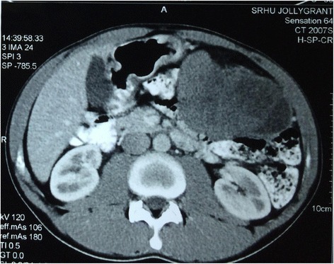
The greater omentum is an uncommon location for primary tumors. Metastatic tumors of the omentum are common. In contrast, primary tumors of the omentum are very rare. Sporadic cases of primary rhabdomyosarcoma (RMS) arising in the abdomen have been reported, but these cases are limited almost exclusively to the pediatric population. We report a well-documented case of primary intra-abdominal RMS in a 21-year-old man who presented with complaints of abdominal pain and lump in left hypochondrium region. Imaging revealed it to be a large mass in the left hypochondrium region displacing the bowel loops. Further investigations revealed omental RMS. The mass had originated from the greater omentum and was excised. Our case is doing well and is presently receiving chemotherapy.
Keywords: Greater omentum; Rhabdomyosarcoma.
Figures


References
-
- Evans KJ, Miller Q, Kline AL. Solid omental tumors. New York: WebMD LLC; 2011. pp. c1994–c2013.
-
- Sigauke E, Rakheja D, Maddox DL, Hladik CL, White CL, Timmons CF, et al. Absence of expression of SMARCB1/INI1 in malignant rhabdoid tumors of the central nervous system, kidneys and soft tissue: an immunohistochemical study with implications for diagnosis. Mod Pathol. 2006;19:717–725. doi: 10.1038/modpathol.3800581. - DOI - PubMed
LinkOut - more resources
Full Text Sources
Other Literature Sources
