. 2015 Sep 14;10(9):e0136926.
doi: 10.1371/journal.pone.0136926. eCollection 2015.

Localized Modeling of Biochemical and Flow Interactions during Cancer Cell Adhesion

Affiliations

Localized Modeling of Biochemical and Flow Interactions during Cancer Cell Adhesion

Julie Behr et al. PLoS One. .

Abstract

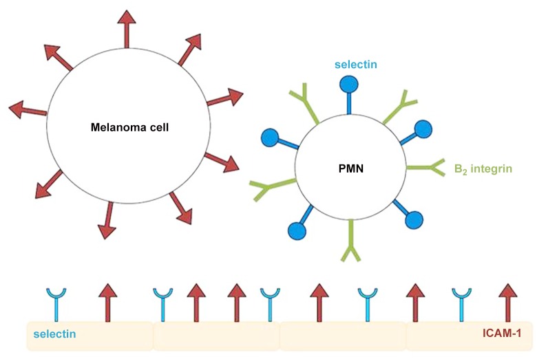
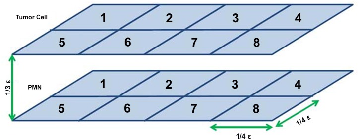
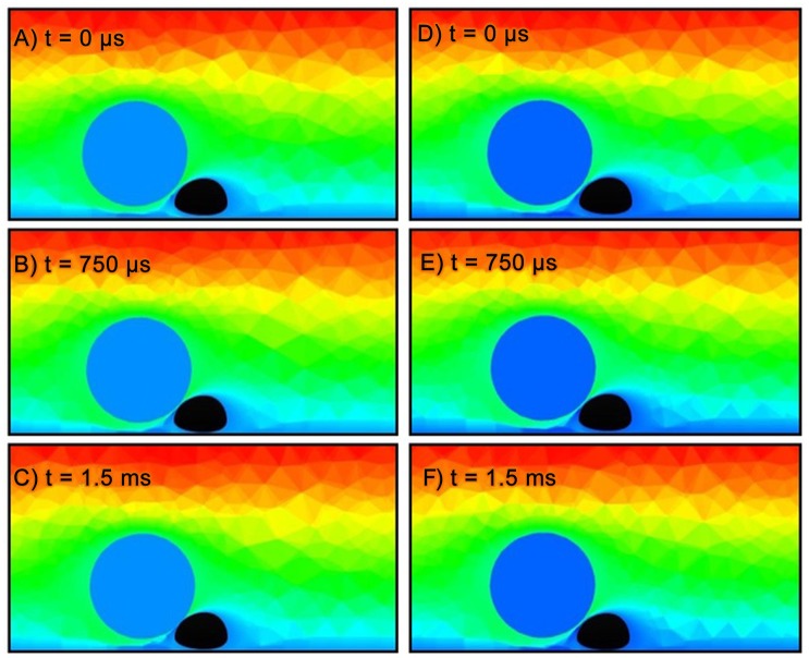
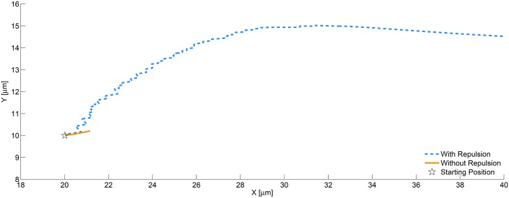
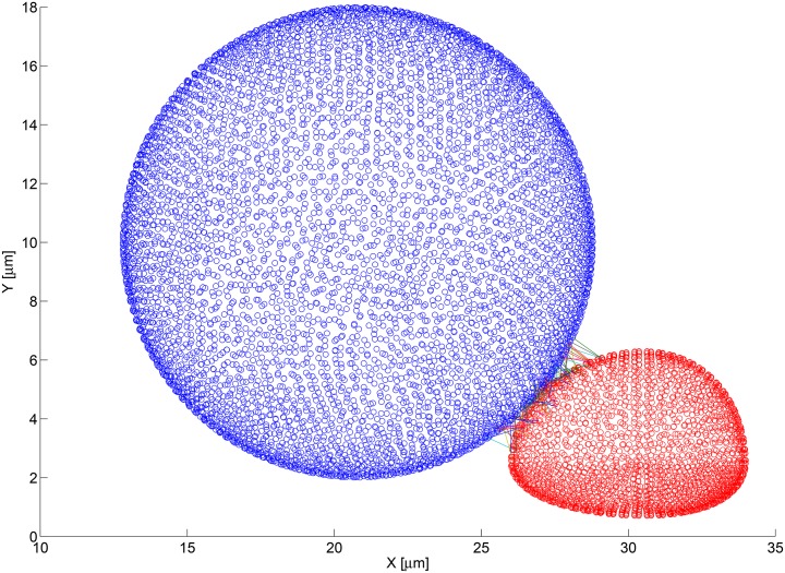
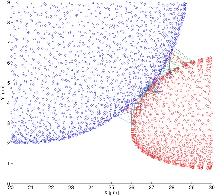
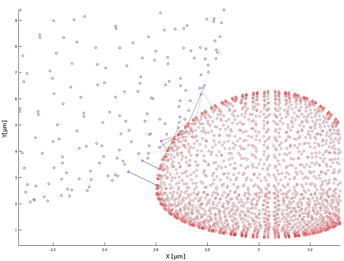
This work focuses on one component of a larger research effort to develop a simulation tool to model populations of flowing cells. Specifically, in this study a local model of the biochemical interactions between circulating melanoma tumor cells (TC) and substrate adherent polymorphonuclear neutrophils (PMN) is developed. This model provides realistic three-dimensional distributions of bond formation and attendant attraction and repulsion forces that are consistent with the time dependent Computational Fluid Dynamics (CFD) framework of the full system model which accounts local pressure, shear and repulsion forces. The resulting full dynamics model enables exploration of TC adhesion to adherent PMNs, which is a known participating mechanism in melanoma cell metastasis. The model defines the adhesion molecules present on the TC and PMN cell surfaces, and calculates their interactions as the melanoma cell flows past the PMN. Biochemical rates of reactions between individual molecules are determined based on their local properties. The melanoma cell in the model expresses ICAM-1 molecules on its surface, and the PMN expresses the β-2 integrins LFA-1 and Mac-1. In this work the PMN is fixed to the substrate and is assumed fully rigid and of a prescribed shear-rate dependent shape obtained from micro-PIV experiments. The melanoma cell is transported with full six-degrees-of-freedom dynamics. Adhesion models, which represent the ability of molecules to bond and adhere the cells to each other, and repulsion models, which represent the various physical mechanisms of cellular repulsion, are incorporated with the CFD solver. All models are general enough to allow for future extensions, including arbitrary adhesion molecule types, and the ability to redefine the values of parameters to represent various cell types. The model presented in this study will be part of a clinical tool for development of personalized medical treatment programs.


Conflict of interest statement

Competing Interests: The authors have declared that no competing interests exist.

Figures

Fig 1
Fig 1. Cartoon of adhesion molecule expression.
β-2 integrins expressed on the PMN can interact with ICAM-1 expressed on both the melanoma cell and endothelial cells. The PMN and endothelium can also interact through selectins.
Fig 2
Fig 2. Simplified computational mesh used for model validation.
This simplified version of a mesh was used to run the Adhesion model through MATLAB without requiring the input of the detailed geometric mesh used by NPHASE.
Fig 3
Fig 3. Comparison of CFD results using same initial parameters, with and without repulsion.
(a-c) show results for the CFD simulation without the cellular repulsion. Without repulsion, the bodies can collide and cause the CFD solver to crash. When a collision occurs, as shown in (c), the CFD may produce non-physical results or be unable to reach a converged solution. (d-f) show results for CFD simulation with cellular repulsion activated. In this case, the bodies do not collide and the simulation continues to advance in time. Note: results are 2D cross-sections of fully 3D simulations. Flow from left to right. Contours are normalized velocity magnitude (blue = 0, red = 1). PMN is colored in black and melanoma cell is colored in blue.
Fig 4
Fig 4. Trajectories of melanoma cell from CFD simulation, with and without repulsion.
The results from the simulation shown in Fig 3 is post-processed to extract the centroid location of the melanoma cell throughout the simulation. That data was used to create a trajectory for each of the two cases. The case without repulsion (solid line) has a short path due to the collision with the PMN cell. The case with repulsion activated (dashed line) shows the body moving over and pass the PMN body. In this particular simulation, the motion of the centroid is planar (z = constant).
Fig 5
Fig 5. Bond formation between nearby cells using the fine computational mesh.
The blue figure is a 3D point cloud of the fine computational mesh used to describe the surface of the melanoma cell. Each blue circle represents the centroid of a discretized mesh face. Similarly, the red figure is a 3D point cloud of the fine computational mesh used to describe the surface of the PMN. The lines connecting the circles represents bonds that have formed and connect the two faces on which the involved adhesion molecules reside.
Fig 6
Fig 6. Zoomed view of bond formation using the fine computational mesh.
This is a zoomed view of the bond formation and 3D point clouds of the PMN and melanoma cell using the fine mesh (shown in Fig 5). A total of 96 bonds were formed and are shown. Note: multiple bonds may occur between the same face pairs.
Fig 7
Fig 7. Zoomed view of bond formation using the coarse computational mesh.
This is a zoomed view of the bond formation and 3D point clouds of the PMN and melanoma cell using the coarse mesh. With the coarse computational mesh, 97 bonds were formed and are shown. Each face pair contains multiple adhesive bonds, which allows for an appropriate number interactions although fewer computational faces are involved in the calculations. Note: multiple bonds may occur between the same face pairs.

References

    1. Dong C, Slattery MJ, Liang S, Peng HH (2005) Melanoma cell extravasation under flow conditions is modulated by leukocytes and endogenously produced interleukin 8. Mol Cell Biomech 2: 145–159. - PMC - PubMed
    1. Slattery MJ, Dong C (2003) Neutrophils influence melanoma adhesion and migration under flow conditions. International journal of cancer 106: 713–722. 10.1002/ijc.11297 - DOI - PMC - PubMed
    1. Di Wu Q, Wang JH, Condron C, Bouchier-Hayes D, Redmond HP (2001) Human neutrophils facilitate tumor cell transendothelial migration. American Journal of Physiology-Cell Physiology 280: C814–C822. - PubMed
    1. Jamali Y, Mohammad A, K MMR (2010) A sub-cellular viscoelastic model for cell population mechanics. PLOS ONE 5: 1–20. 10.1371/journal.pone.0012097 - DOI - PMC - PubMed
    1. Hammer DA, Apte SM (1992) Simulation of cell rolling and adhesion on surfaces in shear flow: general results and analysis of selectin-mediated neutrophil adhesion. Biophysical Journal 63: 35–57. 10.1016/S0006-3495(92)81577-1 - DOI - PMC - PubMed

Publication types