Comparative Study
. 2015 Sep 14;9(9):e0004018.
doi: 10.1371/journal.pntd.0004018. eCollection 2015.

Differential Gene Expression and Infection Profiles of Cutaneous and Mucosal Leishmania braziliensis Isolates from the Same Patient

Affiliations
Comparative Study

Differential Gene Expression and Infection Profiles of Cutaneous and Mucosal Leishmania braziliensis Isolates from the Same Patient

Eliza V C Alves-Ferreira et al. PLoS Negl Trop Dis. .

Abstract

Background: Leishmaniasis is a complex disease in which clinical outcome depends on factors such as parasite species, host genetics and immunity and vector species. In Brazil, Leishmania (Viannia) braziliensis is a major etiological agent of cutaneous (CL) and mucosal leishmaniasis (MCL), a disfiguring form of the disease, which occurs in ~10% of L. braziliensis-infected patients. Thus, clinical isolates from patients with CL and MCL may be a relevant source of information to uncover parasite factors contributing to pathogenesis. In this study, we investigated two pairs of L. (V.) braziliensis isolates from mucosal (LbrM) and cutaneous (LbrC) sites of the same patient to identify factors distinguishing parasites that migrate from those that remain at the primary site of infection.

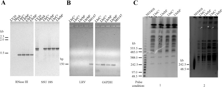
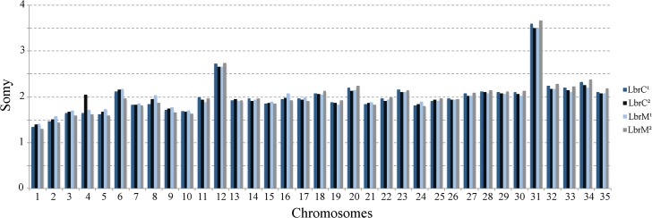
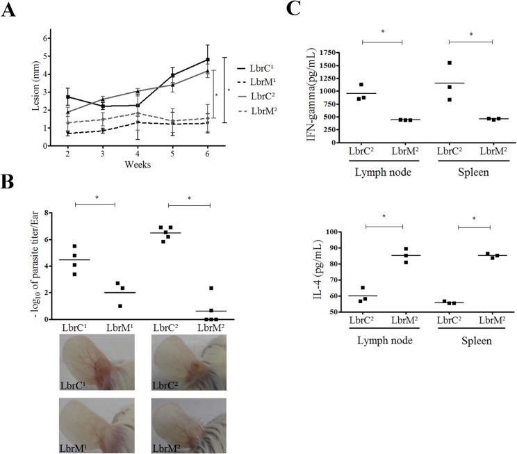
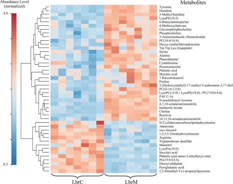
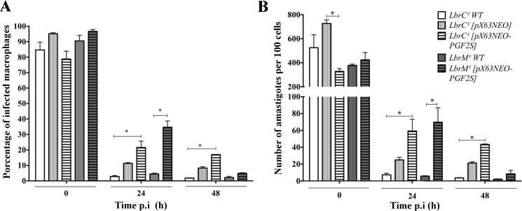
Methodology/principal findings: We observed no major genomic divergences among the clinical isolates by molecular karyotype and genomic sequencing. RT-PCR revealed that the isolates lacked Leishmania RNA virus (LRV). However, the isolates exhibited distinct in vivo pathogenesis in BALB/c mice; the LbrC isolates were more virulent than the LbrM isolates. Metabolomic analysis revealed significantly increased levels of 14 metabolites in LbrC parasites and 31 metabolites in LbrM parasites that were mainly related to inflammation and chemotaxis. A proteome comparative analysis revealed the overexpression of LbrPGF2S (prostaglandin f2-alpha synthase) and HSP70 in both LbrC isolates. Overexpression of LbrPGF2S in LbrC and LbrM promastigotes led to an increase in infected macrophages and the number of amastigotes per cell at 24-48 h post-infection (p.i.).

Conclusions/significance: Despite sharing high similarity at the genome structure and ploidy levels, the parasites exhibited divergent expressed genomes. The proteome and metabolome results indicated differential profiles between the cutaneous and mucosal isolates, primarily related to inflammation and chemotaxis. BALB/c infection revealed that the cutaneous isolates were more virulent than the mucosal parasites. Furthermore, our data suggest that the LbrPGF2S protein is a candidate to contribute to parasite virulence profiles in the mammalian host.


Conflict of interest statement

The authors have declared that no competing interests exist.

Figures

Fig 1
Fig 1. Molecular characterization of the LbrC and LbrM clinical isolates.
(A) Agarose gel-fractionation of the PCR products from DNA extracted from LbrC1, LbrM1, LbrC2 and LbrM2 parasite cultures verified that the isolates belong to the Viannia subgenus. The Viannia subgenus was identified by PCR using primers for the RNase III domain gene (LbrM.23.0390) and 18S rDNA (SSU) gene as a DNA quality control. The LV39 strain [Leishmania (Leishmania) major] and a mock reaction (PCR reaction without genomic DNA) were used as negative controls. (B) PCR analysis of DNA extracted from LbrC1, LbrM1, LbrC2, LbrM2 and M4147 parasite cultures fractionated in agarose gels demonstrated that the Leishmania virus (LRV) was not amplified by RT-PCR from cutaneous and mucosal isolates. cDNA from Leishmania Viannia guyanensis (M4147 strain), which harbors the virus, was used as a positive control, and G6PDH primers were used as a positive control for Leishmania DNA. (C) Molecular karyotypes of parasites rescued from cutaneous and mucosal sites (LbrC1, LbrM1, LbrC2 and LbrM2), as determined by pulsed-field gel electrophoresis (PFGE). The electrophoresis conditions are described in the Methods section. Numbers 1 and 2 refer to electrophoresis programs. Marker: N0340S (NE BioLabs). The gels were stained with ethidium bromide. White arrows indicate bands observed only in the LbrM isolates. * highlights the LbrC2 band with a doubled signal intensity compared to LbrM2.
Fig 2
Fig 2. Chromosome somy of the LbrC and LbrM isolates.
The median coverage for a haploid allele of a chromosome was calculated. The median coverage of each chromosome was divided by the haploid chromosome coverage to obtain the somy of the individual chromosomes [31].
Fig 3
Fig 3. LbrC and LbrM isolates exhibit distinct virulence in BALB/c mice.
(A) The panel represents the lesion development in hamsters (Mesocricetus auratus) after infection with the isolates. Ear thickness was measured with a Mitutuyo digital caliper for six weeks. Each point represents the mean lesion size (±SEM) of five animals per group. (B) The graph represents the parasite load in the ear of BALB/c mice after a four-week infection with the isolates by limiting dilution assay. Three or five mice were used per group. The lower panel shows representative images of ear lesions after 4 weeks of infection. (C) Quantification of IFN-γ and IL-4 cytokines released by lymph node and spleen cells after one month of infection with the LbrC2 and LbrM2 isolates following stimulation with SLA for 72 h. Three mice were included in each group. *p<0.05 (two-tailed t-test).
Fig 4
Fig 4. Hierarchical Clustering Heatmap of biologically relevant metabolites in LbrM and LbrC.
The color code indicates the metabolites abundance. To enable the comparison of data obtained from HPLC-MS, CE-MS and GC-MS the metabolite abundance was normalized. The MetaboAnalyst (v. 2.0) website was used to normalize the data and to generate the figure. The normalization procedure consisted of mean-centering and division by the standard deviation of each variable. The lines in the heatmap represent the relative abundance of metabolites across the samples of the two compared groups, LbrC and LbrM; each metabolite is indicated on the right side of the figure. The columns corresponding to the LbrM and LbrC groups are indicated at the bottom. Each of the seven columns corresponds to one biological replicate (seven per group). To the left-hand side of the figure, a scale indicates the color code relative to the normalized metabolite abundance (ranging from 0.5 up to 4.0).
Fig 5
Fig 5. Ectopic overexpression of LbrPGF2S increases the infection index in vitro.
(A) Peritoneal macrophages from BALB/c mice were infected with Lbrc and LbrM transfectants and the wild type strain. At 0 h, 24 h and 48 h post-infection, the cells were stained, and 600 cells were counted. Each bar represents the average and SD of three replicates. *p<0.05 (Student's t test). (B) Number of amastigotes in the macrophages.

References

    1. Alvar J, Vélez ID, Bern C, Herrero M, Desjeux P, Cano J, et al. Leishmaniasis worldwide and global estimates of its incidence. PLoS One. 2012;7(5):e35671 10.1371/journal.pone.0035671 - DOI - PMC - PubMed
    1. de Almeida MC, Vilhena V, Barral A, Barral-Netto M. Leishmanial infection: analysis of its first steps. A review. Mem Inst Oswaldo Cruz. 2003;98(7):861–70. - PubMed
    1. Teixeira MJ, Teixeira CR, Andrade BB, Barral-Netto M, Barral A. Chemokines in host-parasite interactions in leishmaniasis. Trends Parasitol. 2006;22(1):32–40. - PubMed
    1. Andrade BB, de Oliveira CI, Brodskyn CI, Barral A, Barral-Netto M. Role of sand fly saliva in human and experimental leishmaniasis: current insights. Scand J Immunol. 2007;66(2–3):122–7. - PubMed
    1. Reithinger R, Dujardin JC, Louzir H, Pirmez C, Alexander B, Brooker S. Cutaneous leishmaniasis. Lancet Infect Dis. 2007;7(9):581–96. Epub 2007/08/24. - PubMed

Publication types

LinkOut - more resources