. 2015 Sep 14;10(9):e0138142.
doi: 10.1371/journal.pone.0138142. eCollection 2015.

Sphingosine-1-Phosphate Signaling Regulates Myogenic Responsiveness in Human Resistance Arteries

Affiliations

Sphingosine-1-Phosphate Signaling Regulates Myogenic Responsiveness in Human Resistance Arteries

Sonya Hui et al. PLoS One. .

Abstract

We recently identified sphingosine-1-phosphate (S1P) signaling and the cystic fibrosis transmembrane conductance regulator (CFTR) as prominent regulators of myogenic responsiveness in rodent resistance arteries. However, since rodent models frequently exhibit limitations with respect to human applicability, translation is necessary to validate the relevance of this signaling network for clinical application. We therefore investigated the significance of these regulatory elements in human mesenteric and skeletal muscle resistance arteries. Mesenteric and skeletal muscle resistance arteries were isolated from patient tissue specimens collected during colonic or cardiac bypass surgery. Pressure myography assessments confirmed endothelial integrity, as well as stable phenylephrine and myogenic responses. Both human mesenteric and skeletal muscle resistance arteries (i) express critical S1P signaling elements, (ii) constrict in response to S1P and (iii) lose myogenic responsiveness following S1P receptor antagonism (JTE013). However, while human mesenteric arteries express CFTR, human skeletal muscle resistance arteries do not express detectable levels of CFTR protein. Consequently, modulating CFTR activity enhances myogenic responsiveness only in human mesenteric resistance arteries. We conclude that human mesenteric and skeletal muscle resistance arteries are a reliable and consistent model for translational studies. We demonstrate that the core elements of an S1P-dependent signaling network translate to human mesenteric resistance arteries. Clear species and vascular bed variations are evident, reinforcing the critical need for further translational study.


Conflict of interest statement

Competing Interests: The authors have declared that no competing interests exist.

Figures

Fig 1
Fig 1. Human mesenteric and skeletal muscle resistance artery vasodilator responses.
Human (A) mesenteric (n = 7 vessels from N = 3 patients) and (B) skeletal muscle (n = 8, N = 5) resistance arteries constrict in response to 10-5 mol/L phenylephrine (open circles) and dose-dependently dilate when acetylcholine is subsequently co-applied (closed circles). For mesenteric arteries: diamax = 255±23μm and logEC50 = -7.4±0.2. For skeletal muscle arteries: diamax = 133±22μm and logEC50 = -7.2±0.2. Diamax is defined as the maximal diameter (under calcium free buffer conditions) at 60mmHg.
Fig 2
Fig 2. Response stability in human mesenteric and skeletal muscle resistance arteries.
Time-control experiments confirm that (A) phenylephrine (PE)-stimulated vasoconstriction (logEC50 = -6.4±0.2; diamax = 173±33μm; n = 7 vessels from N = 5 patients), (B) myogenic tone (diamax = 185±34μm; n = 7, N = 4) and (C) active myogenic constriction (following a pressure step from 60mmHg to 100mmHg; diamax = 105±15μm; n = 7, N = 3) are consistent in human mesenteric resistance arteries over two separate assessments. (D) PE-stimulated vasoconstriction (logEC50 = -6.5±0.1; diamax = 104±13μm; n = 10, N = 10) and (E) myogenic tone (diamax = 122±21μm; n = 9, N = 8) are also consistent over two separate assessments in human skeletal muscle resistance arteries; however, (F) active myogenic constriction (diamax = 94±9μm; n = 7, N = 6) is not stable. Diamax is defined as the maximal diameter (under calcium free buffer conditions) at 60mmHg. Myogenic tone (Panels A and D) and PE responses (Panels B and E) were statistically compared with a paired two-way ANOVA; active constriction measures (Panels C and F) were compared with a Wilcoxon test. * denotes P<0.05 for a paired comparisons.
Fig 3
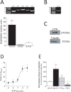
Fig 3. S1P signaling in human mesenteric resistance arteries.
(A) Human mesenteric resistance arteries express mRNA encoding critical sphingosine-1-phosphate (S1P) signaling elements (N = 8 patients), including sphingosine kinase 1 (Sphk1), S1P phosphohydrolase 1 (SPP1) and S1P receptor subtypes 1–3 (S1P1R, S1P2R and S1P3R). In terms of relative expression, S1P1R is the most highly-expressed receptor, followed by S1P3R and then S1P2R (N = 8). Human mesenteric arteries express the cystic fibrosis transmembrane conductance regulator (CFTR) at both the (B) mRNA (N = 8) and (C) protein levels (N = 3); in (C), tubulin confirms adequate loading. (D) S1P stimulates dose-dependent vasoconstriction (logEC50 = -6.7±0.2; diamax = 121±13μm; n = 5 vessels from N = 3 patients). (E) Shown are normalized active constriction measurements (post-treatment measures normalized to pre-treatment responses). At sub-threshold concentrations (i.e., levels that do not stimulate overt constriction), S1P dose-dependently enhances myogenic vasoconstriction (1nmol/L S1P: diamax = 177±27μm, n = 7, N = 5; 10nmol/L S1P: diamax = 165±19μm, n = 11, N = 6); CFTR inhibition (1nmol/L CFTR(inh)-172: diamax = 203±13μm, n = 12, N = 6) also enhances myogenic vasoconstriction, while S1P receptor antagonism (10nmol/L JTE013: diamax = 148±30μm, n = 7, N = 4) attenuates myogenic vasoconstriction. Diamax is defined as the maximal diameter (under calcium free buffer conditions) at 60mmHg. In Panel A, * denotes P<0.05 for S1P1R relative to the other two receptors (Friedman ANOVA); in Panel E, * denotes P<0.05 for a paired comparison to the pre-treatment response (Wilcoxon test).
Fig 4
Fig 4. S1P signaling in human skeletal muscle resistance arteries.
(A) Human skeletal muscle resistance arteries express mRNA encoding critical sphingosine-1-phosphate (S1P) signaling elements (N = 8 patient samples), including sphingosine kinase 1 (Sphk1), S1P phosphohydrolase 1 (SPP1) and S1P receptor subtypes 1–3 (S1P1R, S1P2R and S1P3R). In terms of relative expression, S1P1R is the most highly expressed; S1P3R is more highly expressed than S1P2R (N = 8). Human skeletal muscle resistance arteries (B) express detectable levels of cystic fibrosis transmembrane conductance regulator (CFTR) mRNA (N = 8); however, (C) CFTR protein is not detected (N = 5). In (C), tubulin confirms adequate loading. (D) S1P stimulates dose-dependent vasoconstriction (diamax = 122.2±16.5μm; n = 6 vessels from N = 4 patients). (E) S1P receptor antagonism (100pmol/L JTE013: diamax = 132±15μm, n = 8, N = 5) attenuates myogenic tone. However, neither (F) sub-threshold concentrations of S1P (10nmol/L S1P: diamax = 110±20μm; n = 5, N = 4) nor (G) CFTR inhibition (10pmol/L CFTR(inh)-172: diamax = 139±33μm, n = 5, N = 4) modulate myogenic tone. Diamax is defined as the maximal diameter (under calcium free buffer conditions) at 60mmHg. In Panel A, * denotes P<0.05 relative to all other receptor subtypes (Friedman ANOVA). Panels E, F and G were statistically compared with a paired two-way ANOVA; in Panel E, * denotes P<0.05 relative to the pre-treatment control response.

References

    1. Braun O, Insel PA. The Best “Model System” for Human (Coronary Arteries) Is Human. Journal of the American College of Cardiology. 2009;54: 1146–1148. 10.1016/j.jacc.2009.06.019 - DOI - PubMed
    1. Jensen BC, Swigart PM, Laden ME, DeMarco T, Hoopes C, Simpson PC. The Alpha-1D Is the Predominant Alpha-1-Adrenergic Receptor Subtype in Human Epicardial Coronary Arteries. Journal of the American College of Cardiology. 2009;54: 1137–1145. 10.1016/j.jacc.2009.05.056 - DOI - PMC - PubMed
    1. Paulson OB, Strandgaard S, Edvinsson L. Cerebral autoregulation. Cerebrovasc Brain Metab Rev. 1990;2: 161–192. - PubMed
    1. Just A. Mechanisms of renal blood flow autoregulation: dynamics and contributions. Am J Physiol Regul Integr Comp Physiol. 2007;292: R1–17. - PubMed
    1. Metting PJ, Kostrzewski KA, Stein PM, Stoos BA, Britton SL. Quantitative contribution of systemic vascular autoregulation in acute hypertension in conscious dogs. J Clin Invest. 1989;84: 1900–1905. - PMC - PubMed

Publication types