. 2015 Sep 14;10(9):e0137837.
doi: 10.1371/journal.pone.0137837. eCollection 2015.

Pycnogenol Attenuates the Release of Proinflammatory Cytokines and Expression of Perilipin 2 in Lipopolysaccharide-Stimulated Microglia in Part via Inhibition of NF-κB and AP-1 Activation

Affiliations

Pycnogenol Attenuates the Release of Proinflammatory Cytokines and Expression of Perilipin 2 in Lipopolysaccharide-Stimulated Microglia in Part via Inhibition of NF-κB and AP-1 Activation

Bin Fan et al. PLoS One. .

Abstract

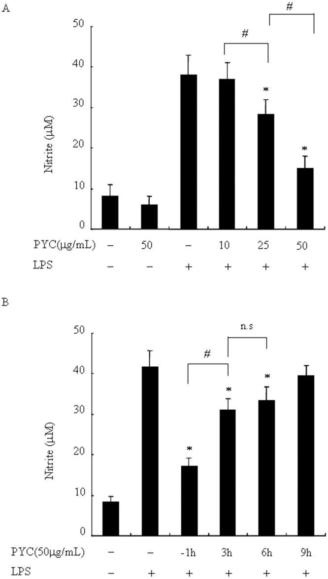
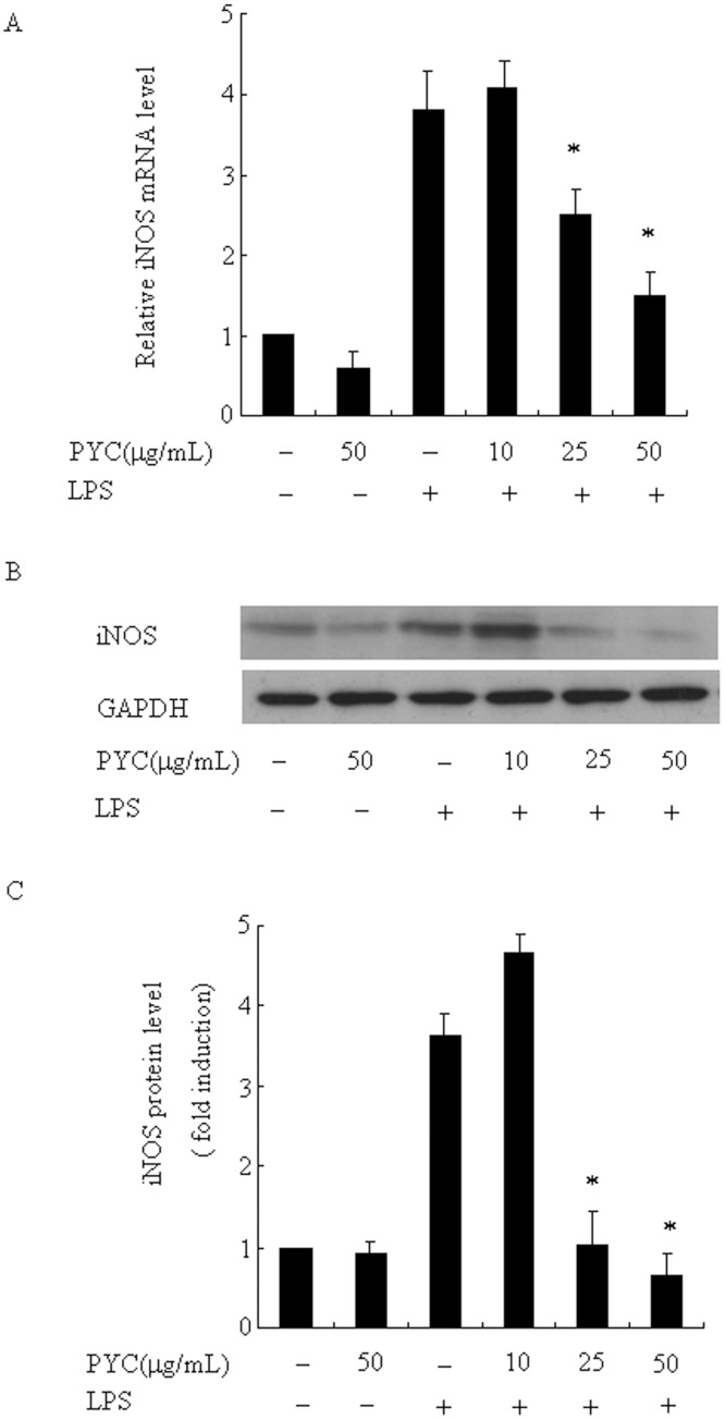
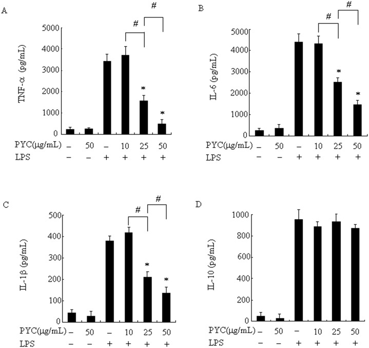
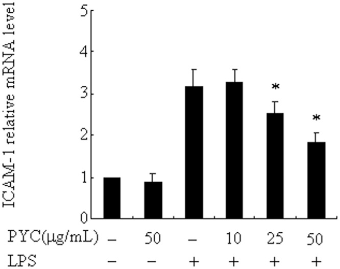
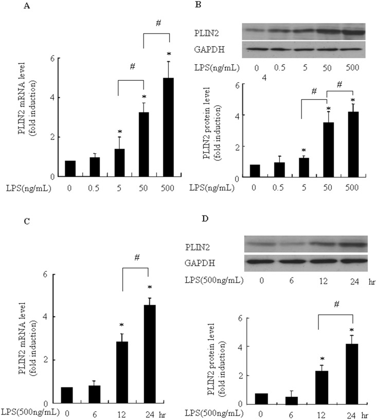
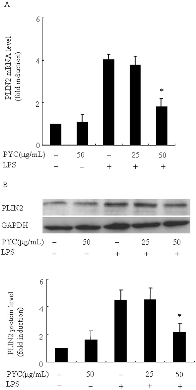
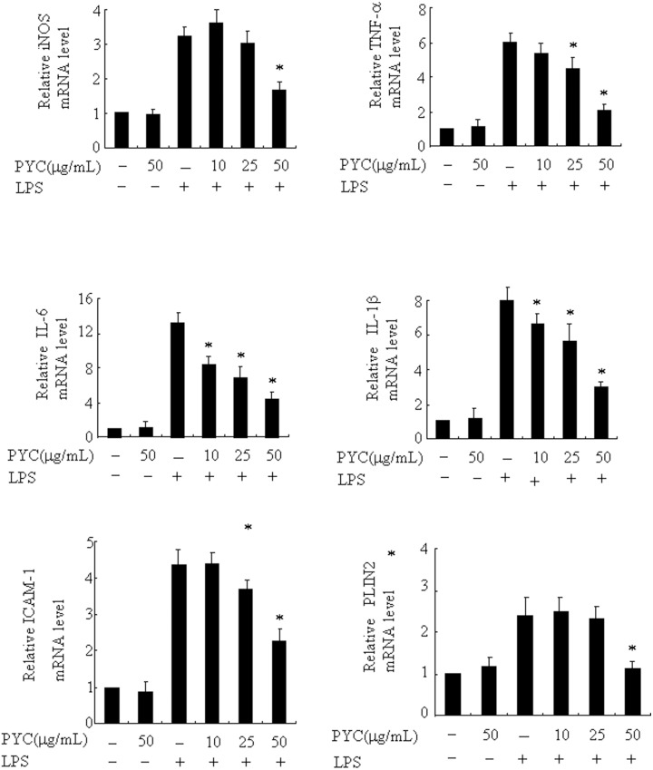
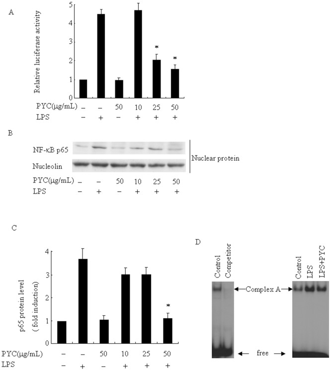
Over activation of microglia results in the production of proinflammatory agents that have been implicated in various brain diseases. Pycnogenol is a patented extract from French maritime pine bark (Pinus pinaster Aiton) with strong antioxidant and anti-inflammatory potency. The present study investigated whether pycnogenol may be associated with the production of proinflammatory mediators in lipopolysaccharide-stimulated BV2 (mouse-derived) microglia. It was found that pycnogenol treatment was dose-dependently associated with significantly less release of nitricoxide (NO), TNF-α, IL-6 and IL-1β, and lower levels of intercellular adhesion molecule1 (ICAM-1) and perilipin 2 (PLIN2). Furthermore, this effect was replicated in primary brain microglia. Levels of inducible NO synthase mRNA and protein were attenuated, whereas there was no change in the production of the anti-inflammatory cytokine IL-10. Further evidence indicated that pycnogenol treatment led to the suppression of NF-κB activation through inhibition of p65 translocation into the nucleus and inhibited DNA binding of AP-1, suggesting that these proinflammatory factors are associated with NF-κB and AP-1. We conclude that pycnogenol exerts anti-inflammatory effects through inhibition of the NF-κB and AP-1pathway, and may be useful as a therapeutic agent in the prevention of diseases caused by over activation of microglia.


Conflict of interest statement

Competing Interests: The authors have declared that no competing interests exist.

Figures

Fig 1
Fig 1. PYC suppressed LPS-induced NO production in BV2 microglia.
Cells were incubated with PYC (10, 25, 50 μg/mL) or DMSO (vehicle) for 1 h before addition of LPS (500ng/mL) for 24h (A) or cells were incubated with PYC (50 μg/mL) or vehicle in indicated time before or after LPS stimulation for 24h (B). The culture supernatants were collected and analyzed for nitrite production using Griess reagent and a standard curve was created using NaNO2 in culture medium. Data are represented as mean ± SD from at least 3 independent experiments. *P< 0.05 compared with LPS alone. #P< 0.05.
Fig 2
Fig 2. Effects of different doses of PYC on cell viability of BV2 microglia.
BV2 cells were treated with PYC at various concentrations or DMSO (vehicle) for 1h. Then, LPS (500ng/mL) was added and incubated for 24h. After treatments, Cell viability was assessed by MTT and the results are expressed as the percentage of surviving cells over control cells (no addition of PYC and LPS). Data are represented as mean ± SD from at least 3 independent experiments.
Fig 3
Fig 3. Effects of different doses of PYC on LPS-induced the levels of iNOS mRNA and protein in BV2 microglia.
BV2 cells were treated with PYC at various concentrations or DMSO (vehicle) (10, 25, 50 μg/mL) for 1h. LPS (500ng/mL) was then added and incubated for an additional 24 h. Ribosomal RNAs and GAPDH were used as the total RNA or protein loading control, respectively. (A) iNOS mRNA was assessed by real-time PCR, and the mRNA level in the control (no stimuli) was arbitrarily designated as 1 for comparison. The data represent the means ± SD of 3 independent experiments. *P< 0.05 compared with LPS alone. (B) Levels of iNOS protein were assessed via Western blot. (C) Levels of iNOS protein were quantified by the NIH Image processing and analysis program. *P< 0.05 compared with LPS alone. Experiments were repeated at least 3 times and similar results were obtained.
Fig 4
Fig 4. PYC suppressed LPS-induced proinflammatory cytokine production in BV2 microglia.
Cells were incubated with the indicated concentrations of PYC or vehicle for 1h before LPS treatment (500ng/mL). After 24 h incubation, the culture supernatants were collected, and the amount of TNF-α, IL-6, IL-1β and IL-10 were measured by ELISA (A-D). Data are represented as mean ± SD from at least 3 independent experiments. *P< 0.05 compared with LPS alone. #P< 0.05.
Fig 5
Fig 5. Pre-incubation of different doses PYC suppressed LPS-induced ICAM-1 expression in BV2 microglia.
BV2 cells were treated with PYC or vehicle at the indicated concentrations for 1h. LPS (500ng/mL) was then added and further incubated for 24 h. Ribosomal RNAs were used as the total RNA loading control. ICAM-1 mRNA was assessed by real-time PCR, and the mRNA level in the control (no stimuli) was arbitrarily designated as 1 for comparison. The data represent the means ± SD of at least 3 independent experiments. *P< 0.05 compared with LPS alone.
Fig 6
Fig 6. LPS increased the levels of PLIN2 mRNA and protein in a dose and time dependent manner in BV2 microglia.
(A and B): LPS stimulated PLIN2 mRNA and protein expression in a dose-dependent manner. Cells were incubated with vehicle or indicated concentrations of LPS for 24 h. (C and D): LPS enhanced PLIN2 mRNA and protein expression in a time-dependent manner. Cells were incubated with vehicle or LPS (500ng/mL) for 6, 12 or 24h. Ribosomal RNAs and GAPDH were used as the total RNA or protein loading control, respectively. The PLIN2 mRNA level in the control (no stimuli) was arbitrarily designated as 1 for comparison. Levels of PLIN2 protein were quantified by the NIH Image processing and analysis program. *P< 0.05 compared with LPS alone. #P< 0.05. Experiments were repeated 3 times and representative results are shown.
Fig 7
Fig 7. Pre-incubation of different doses PYC suppressed LPS-induced PLIN2 expression in BV2 microglia.
Cells were incubated with the indicated concentrations of PYC or vehicle for 1h before 24 h LPS treatment (500ng/mL). Ribosomal RNAs and GAPDH were used as the total RNA or protein loading control, respectively. (A) PLIN2 mRNA was assessed by real-time PCR, and the mRNA level in the control (no stimuli) was arbitrarily designated as 1 for comparison. (B) Levels of PLIN2 protein were assessed via Western blot. Levels of PLIN2 protein were quantified by the NIH Image processing and analysis program. *P< 0.05 compared with LPS alone. #P< 0.05. Representative data are shown. Relative mRNA and protein levels obtained from 3 independent experiments are shown in bar graphs.
Fig 8
Fig 8. The inhibitory potential of PYC was also fully replicated in mouse primary brain microglia.
Cells were treated with PYC or vehicle at the indicated concentrations for 1h. LPS (500ng/mL) was then added and further incubated for 24 h. Ribosomal RNAs were used as the total RNA loading control. mRNA levels was assessed by real-time PCR, and the mRNA level in the control (no stimuli) was arbitrarily designated as 1 for comparison. The data represent the means ± SD of at least 3 independent experiments. *P< 0.05 compared with LPS alone.
Fig 9
Fig 9. PYC suppressed LPS-induced NF-κB activity and DNA binding of AP-1 in BV2 microglia.
(A) NF-κB-luciferase reporter plasmids were transfected in BV2 cells for 4 h, pre-treated with PYC 10, 25, and 50 μg/mL or DMSO (vehicle) for 1 h, then challenged with 500ng/mL LPS for 6 h. NF-κB activity was expressed as relative luciferase activity. *P< 0.05 compared with LPS alone. Cells were treated with indicated doses of PYC or DMSO (vehicle) for 1 h before 500ng/mL LPS treatment for 30 min, and then subjected to Western blot to detect p65 protein levels (B) and EMSA to assess AP-1 activation (C). 32P-end-labeled AP1 consensus oligonucleotide was used as a probe. Unlabeled oligonucleotide was used as a competitor. The experiment was repeated at least 3 times and similar results were obtained.
Fig 10
Fig 10. The effects of the combination of PYC and NF-κB inhibitor (PDTC) on TNF-α and PLIN2 mRNA levels.
Cells were treated with PYC, PDTC (50, 100 μM) or their combination of both for 1h in LPS (500 ng/mL) was then added and further incubated for 24 h. Ribosomal RNAs were used as the total RNA loading control. mRNA level was assessed by real-time PCR, and the mRNA level in the control (no stimuli) was arbitrarily designated as 1 for comparison. The data represent the means ± SD of at least 3 independent experiments. *P< 0.05 compared with LPS alone.

References

    1. Dang Y, Xu Y, Wu W, Li W, Sun Y, Yang J, et al. Tetrandrine suppresses lipopolysaccharide-induced microglial activation by inhibiting NF-kappaB and ERK signaling pathways in BV2 cells. PloS one. 2014;9(8):e102522 10.1371/journal.pone.0102522 - DOI - PMC - PubMed
    1. Giulian D, Li J, Li X, George J, Rutecki PA. The impact of microglia-derived cytokines upon gliosis in the CNS. Dev Neurosci. 1994;16(3–4):128–36. Epub 1994/01/01. . - PubMed
    1. Lei B, Mace B, Dawson HN, Warner DS, Laskowitz DT, James ML. Anti-inflammatory effects of progesterone in lipopolysaccharide-stimulated BV-2 microglia. PloS one. 2014;9(7):e103969 10.1371/journal.pone.0103969 - DOI - PMC - PubMed
    1. Meda L, Cassatella MA, Szendrei GI, Otvos L Jr, Baron P, Villalba M, et al. Activation of microglial cells by beta-amyloid protein and interferon-gamma. Nature. 1995;374(6523):647–50. Epub 1995/04/13. 10.1038/374647a0 . - DOI - PubMed
    1. Block ML, Hong JS. Microglia and inflammation-mediated neurodegeneration: multiple triggers with a common mechanism. Prog Neurobiol. 2005;76(2):77–98. Epub 2005/08/06. S0301-0082(05)00067-5 [pii] 10.1016/j.pneurobio.2005.06.004 . - DOI - PubMed

Publication types

MeSH terms