. 2015 Nov 1;98(1):132-43.
doi: 10.1016/j.bcp.2015.08.109. Epub 2015 Sep 12.

Toll-like receptor 4 signaling: A common pathway for interactions between prooxidants and extracellular disulfide high mobility group box 1 (HMGB1) protein-coupled activation

Affiliations

Toll-like receptor 4 signaling: A common pathway for interactions between prooxidants and extracellular disulfide high mobility group box 1 (HMGB1) protein-coupled activation

Yan Zhang et al. Biochem Pharmacol. .

Abstract

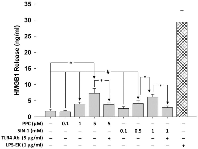
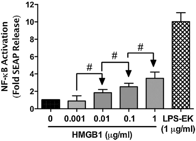
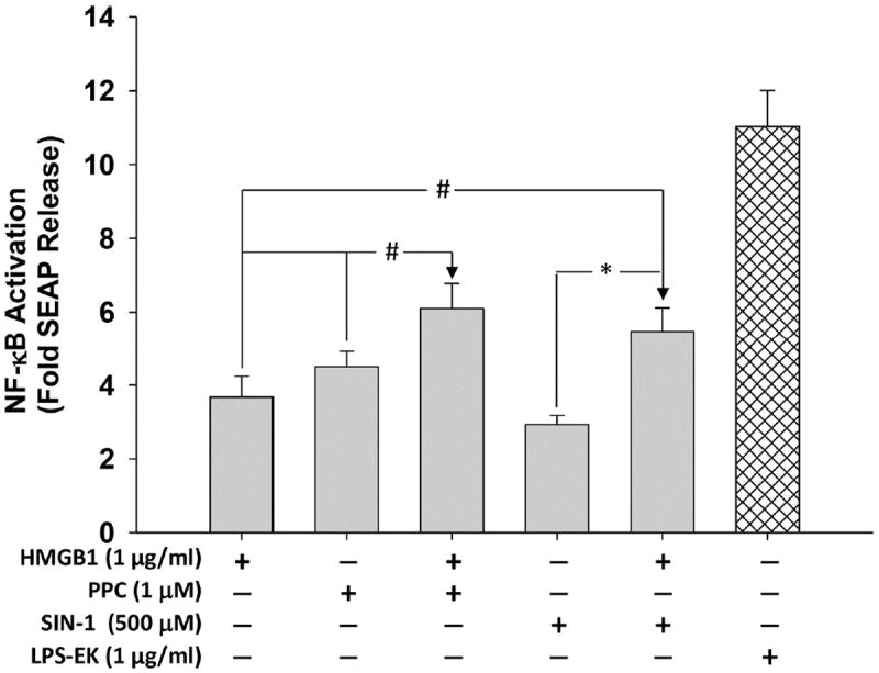
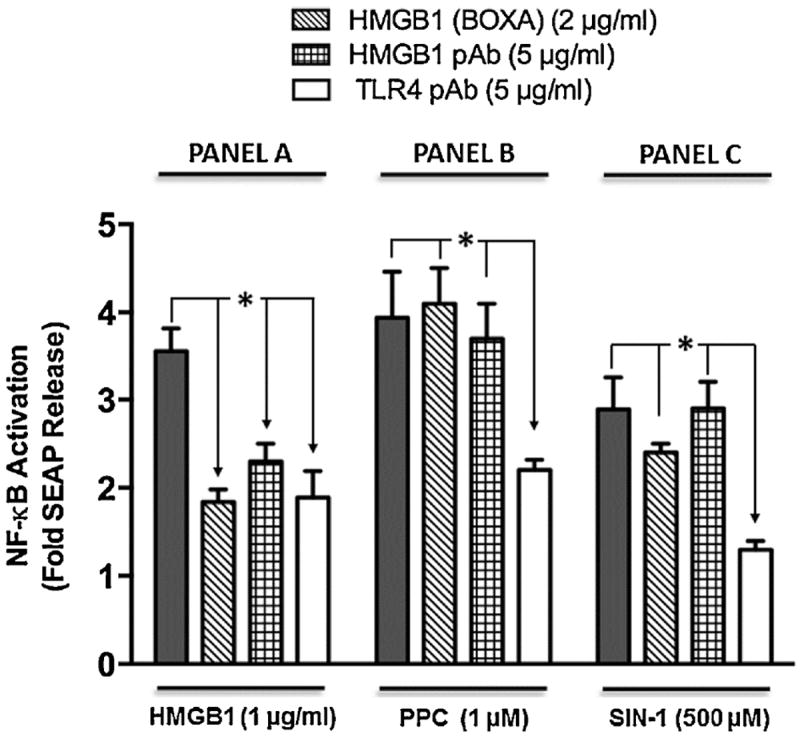
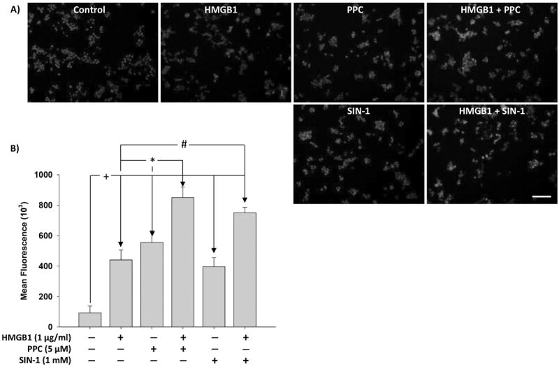
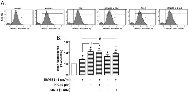
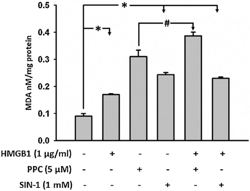
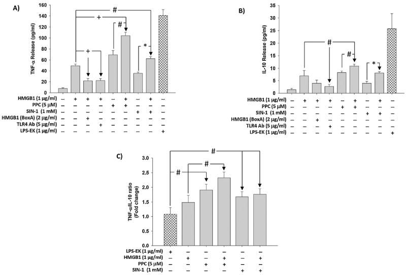
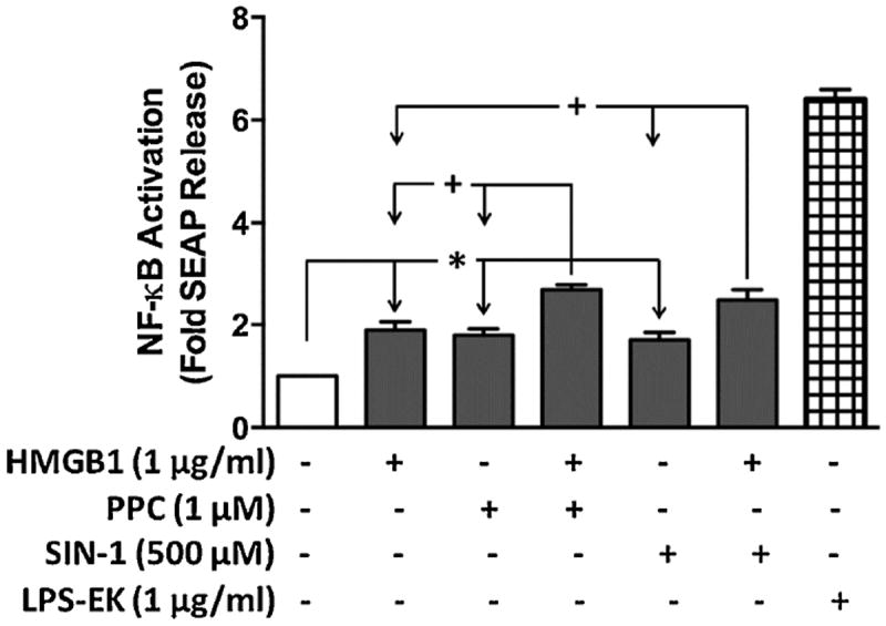
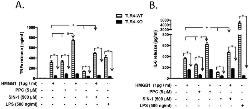
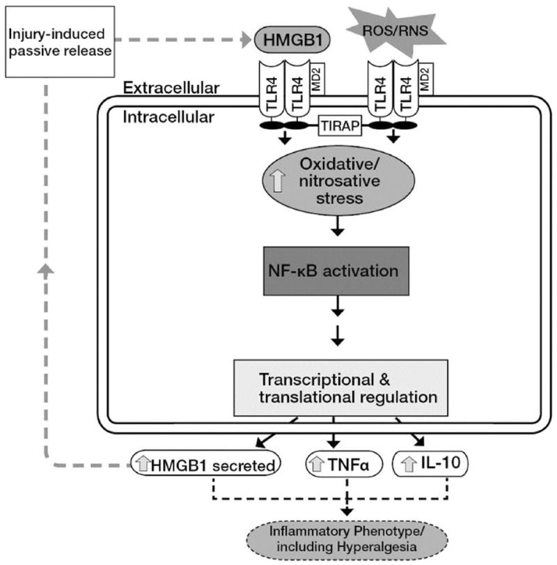
Necrotic cells passively release HMGB1, which can stimulate TLR4 in an autocrine fashion to potentially initiate "sterile" inflammation that maintains different disease states. We have shown that prooxidants can induce NF-κB activation through TLR4 stimulation. We examined whether prooxidants enhance HMGB1-induced TLR4 signaling through NF-κB activation. We used LPS-EK as a specific agonist for TLR4, and PPC and SIN-1 as in situ sources for ROS. As model systems, we used HEK-Blue cells (stably transfected with mouse TLR4), RAW-Blue™ cells (derived from murine RAW 264.7 macrophages) and primary murine macrophages from TLR4-KO mice. Both HEK-Blue and RAW-Blue 264.7 cells express optimized secreted embryonic alkaline phosphatase (SEAP) reporter under the control of a promoter inducible by NF-κB. We treated cells with HMGB1 alone and/or in conjunction with prooxidants and/or inhibitors using SEAP release as a measure of TLR4 stimulation. HMGB1 alone and/or in conjunction with prooxidants increased TNFα and IL-6 released from TLR4-WT, but not from TLR4-KO macrophages. Pro-oxidants increased HMGB1 release, which we quantified by ELISA. We used both fluorescence microscopy imaging and flow cytometry to quantify the expression of intracellular ROS. TLR4-neutralizing antibody decreased prooxidant-induced HMGB1 release. Prooxidants promoted HMGB1-induced NF-κB activation as determined by increased release of SEAP and TNF-α, and accumulation of iROS. HMGB1 (Box A), anti-HMGB1 and anti-TLR4-neutralizing pAbs inhibited HMGB1-induced NF-κB activation, but HMGB1 (Box A) and anti-HMGB1 pAb had no effect on prooxidant-induced SEAP release. The present results confirm that prooxidants enhance proinflammatory effects of HMGB1 by activating NF-κB through TLR4 signaling.

Keywords: NF-κB activation; Prooxidants; Recombinant high mobility group box 1 protein; Sterile inflammation; Toll-like receptor 4.


Conflict of interest statement

The authors have no conflict of interest.

Figures

Fig. 1
Fig. 1. Effect of pro-oxidants on disulfide HMGB1 isoform release in HEK-Blue mTLR4 cells
Cells were treated overnight (~16 h) with varying concentrations of PPC or SIN-1. HMGB1 released into the conditioned media was determined using HMGB1 ELISA kit according to the manufacturer’s instructions. The effect of LPS-EK on HMGB1 release was used as positive control. The data represent 3 independent experiments carried out in duplicate, #p ≤ 0.05 and *p ≤ 0.01.
Fig. 2
Fig. 2. Effect of disulfide HMGB1 isoform on secreted embryonic alkaline phosphatase (SEAP) release in HEK-Blue mTLR4 cells
Cells were treated overnight with varying concentrations of HMGB1. SEAP released into the conditioned medium was determined using Quanti-Blue, and the absorbance was read at 650 nm. The effect of LPS-EK on SEAP release was used as positive control. The data represent 5 independent experiments carried out in duplicate. #p ≤ 0.05
Fig. 3
Fig. 3. Effect of co-treatment of disulfide HMGB1 and prooxidants on secreted embryonic alkaline phosphatase (SEAP) release in HEK-Blue mTLR4 cells
Cells were treated with HMGB1, PPC and SIN-1 alone and/or in combination for 16 h. SEAP released into the conditioned medium was determined using Quanti-Blue according to the manufacturer’s instructions, and the absorbance was read at 650 nm. The effect of LPS-EK on SEAP release was used as positive control. The data represent 5 independent experiments carried out in duplicate. #p ≤ 0.05 and *p ≤ 0.01
Fig. 4
Fig. 4. Effect of TLR4 neutralizing polyclonal antibody (TLR4 pAb), HMGB1 pAb and HMGB1 (Box A) on SEAP release in HEK-Blue mTLR4
Cells were pre-incubated with TLR4 neutralizing antibody, HMGB pAb or HMGB1 (BoxA) for 2 h followed by treatment overnight with HMGB1 (Panel A), PPC (Panel B) or SIN-1 (Panel C) in the continued presence of TLR4 pAb, HMGB1 pAb or HMGB1 (BoxA). SEAP released into the conditioned medium was determined using Quanti-Blue™ with absorbance read at 650 nm. The data represent 3 - 7 independent experiments that were carried out in duplicate. +p ≤ 0.001
Fig. 5
Fig. 5. Immunofluorescence representation of the levels of intracellular ROS (iROS) accumulated following stimulation of HEK-Blue mTLR4 cells
Cells were treated overnight with HMGB1, PPC or SIN-1 alone and/or in combination. Cells were then stained with CellRox® Deep Red Reagent and NucBlue® Live ReadyProbes® Reagent (to counter stain cellular nuclei) for 30 min at 37 °C followed by subsequent PBS washes and fixation with 4 % paraformaldehyde for 15 min. After subsequent rinses with PBS, images were acquired using fluorescence microscope and analyzed using the ImageJ® software. (Fig. 5A) are merged representative pictures with scale bar = 50 μm. (Fig. 5B) represents semiquantitative histograms generated using ImageJ® software. The data represent 3 independent experiments carried out in duplicate. #p ≤ 0.05, *p ≤ 0.01 and +p ≤ 0.001.
Fig. 6
Fig. 6. Flow cytometric analysis of intracellular ROS (iROS) accumulation in HEK-Blue mTLR4 cells following different treatment
Cells were treated overnight with HMGB1, PPC or SIN-1 alone and/or in combination. Following the removal of the incubation media, cells were incubated with CellRox® Deep Red Reagent for 30 min. Cells were rinsed with PBS, then scraped and resuspended into warm PBS according to the manufacturer’s instructions for flow cytometric analysis. Representative tracings (Fig 6A) following treatments with media (control), disulfide HMGB1, PPC or SIN-1 alone or in combination i.e., [HMGB + PPC] or [HMGB1 + SIN-1]. Quantitative analyses of tracings from (Fig. 6A) show changes in fluorescence intensity after different treatments (Fig. 6B). The data in Fig. 6B represent multiple independent experiments conducted in duplicate (n = 5-6; #p ≤ 0.05; *p ≤ 0.05).
Fig. 7
Fig. 7. Levels of malonyldialdehyde (MDA) quantified as thiobarbituric acid reactive substances (TBARS) following stimulation of HEK-Blue mTLR4 cells
Cells were stimulated overnight with HMGB1, PPC or SIN-1 alone and/or in combination in continuous presence of the stimulators. At the end of incubation, the conditioned media was removed and cells rinsed with phosphate buffered saline (PBS). Attached cells were scraped, lysed, freeze thawed once, and centrifuged. The lysates were used to quantify MDA as TBARS according to the manufacturer’s instructions. The data represent 3 independent experiments carried out in duplicate (#p = 0.5, *p ≤ 0.01).
Fig. 8
Fig. 8. Effect of disulfide HMGB1 alone and /or in combination with prooxidants on TNF-α and IL-10 release in HEK-Blue mTLR4 cells
Cells were pre-incubated with HMGB1 (BoxA) or TLR4 pAb followed by stimulation overnight with HMGB1 in continued presence of the inhibitors. In the same set of experiments, cells were also incubated overnight with HMGB1, PPC or SIN-1 alone and/or in combination. Levels of TNF-α (Fig. 8A) and IL-10 (Fig. 8B) released into the culture medium were quantified using the ELISA kits of the relevant cytokine according to manufacturer’s instructions. The ratios of TNF-α to IL-10 (Fig. 8C) were calculated. The effect of LPS-EK on TNF-α and IL-10 release was used as positive control. The data represent 3 independent experiments. (#p ≤ 0.05, *p ≤ 0.01 and +p ≤ 0.001).
Fig. 9
Fig. 9. The effect of prooxidants or HMGB1 alone and /or in combination on SEAP release in RAW-Blue™ macrophage cells that express multiple TLRs (except TLR5)
RAW-Blue™ cells are useful as TLR reporter cells for induction of NF-κB, with a subsequent release of SEAP after treatment with appropriate TLR-specific ligands, including ROS for TLR4 [23]. The QUANTI-Blue™ detection system was used to provide an easy and rapid means to quantify SEAP released into the culture media (based on the extent of TLR4 stimulation). *p ≤ 0.01 and +p ≤ 0.001 with n = 3 independent experiments conducted in duplicate.
Fig. 10
Fig. 10. Effect of HMGB1 alone and /or in combination with prooxidants on TNF-α and IL-6 release in primary murine macrophages derived from TLR4-WT and TLR4-KO mice
Primary macrophages derived from TLR4-WT and TLR4-KO mice were incubated overnight with disulfide HMGB1, PPC or SIN-1 alone and/or in combination as HMGB1+PPC] and HMGB1+SIN-1]. In all cases of [HMGB1+prooxidant] treatments, cells were preincubated with HMGB1 for 1h followed by overnight stimulation with either PPC or SIN-1 in the continued presence of HMGB1. The levels of TNF-α (Fig. 10A) and IL-6 (Fig. 10B) released into the culture media were quantified using the ELISA kits of the relevant murine cytokine according to manufacturer’s instructions. The effect of LPS-EK on TNF-α and IL-6 release was used as positive control. The data represent 3 - 6 independent experiments conducted in duplicate with *p < 0.0001; +p ≤ 0.01; #p ≤ 0.001, PPC or HMGB1 in TLR4-WT compared with [HMGB1 + PPC] combination; +p < 0.01, SIN-1 or HMGB1 in TLR4-WT compared with [HMGB1 + SIN-1] combination.
Fig. 11
Fig. 11. A simplified schematic representation of a putative mechanism for injury-induced exogenous prooxidant-coupled HMGB1 stimulation of TLR4 signaling through NFκB activation
Prooxidants and HMGB1 act in concert to stimulate TLR4. Extracellularly released HMGB1 due to injury can potentially activate TLR4 in an autocrine or paracrine fashion. TLR4 is stimulated in response to exogenous oxidants resulting in the activation of c-Src, which interacts with TLR4 [32]. Increased formation of TLR4/c-Src complex leads to enhanced recruitment of different cytosolic adaptor proteins including toll-interleukin 1 receptor adaptor protein (TIRAP) that results in c-Src/NFκB/IκBα coupled activation.

Similar articles

Cited by

References

    1. Davies KJ. Oxidative stress: the paradox of aerobic life. Biochem Soc Symp. 1995;61:1–31. - PubMed
    1. Giustarini D, Dalle-Donne I, Tsikas D, Rossi R. Oxidative stress and human diseases: Origin, link, measurement, mechanisms, and biomarkers. Crit Rev Clin Lab Sci. 2009;46(5-6):241–281. - PubMed
    1. Davies KJ. Oxidative stress, antioxidant defenses, and damage removal, repair, and replacement systems. IUBMB Life. 2000;50(4-5):279–289. Review. - PubMed
    1. Gill R, Tsung A, Billiar T. Linking oxidative stress to inflammation: Toll-like receptors. Free Radic Biol Med. 2010;48(9):1121–1132. - PMC - PubMed
    1. Xiang M, Fan J, Fan J. Association of Toll-like receptor signaling and reactive oxygen species: a potential therapeutic target for post-trauma acute lung injury. Mediators Inflamm. 2010 doi: 10.1155/2010/916425. - DOI - PMC - PubMed