. 2015 Nov;12(11):1627-37.
doi: 10.1513/AnnalsATS.201507-463OC.

Effect of Antiretroviral Therapy on HIV-mediated Impairment of the Neutrophil Antimycobacterial Response

Affiliations

Effect of Antiretroviral Therapy on HIV-mediated Impairment of the Neutrophil Antimycobacterial Response

David M Lowe et al. Ann Am Thorac Soc. 2015 Nov.

Abstract

Rationale: Experimental and epidemiological evidence suggests that neutrophils are important in the host response to tuberculosis. HIV infection, which increases the risk of tuberculosis, adversely affects neutrophil function.

Objectives: To determine the impact of HIV and antiretroviral therapy on neutrophil antimycobacterial activity.

Methods: We performed a cross-sectional comparison of neutrophil functions in 20 antiretroviral-naive HIV-infected and 20 HIV-uninfected individuals using luminescence-, flow cytometry-, and ELISA-based assays. We then conducted a prospective study in the HIV-infected individuals investigating these parameters during the first 6 months of antiretroviral therapy. Surface markers of neutrophil activation were investigated in a separate cohort using flow cytometry.

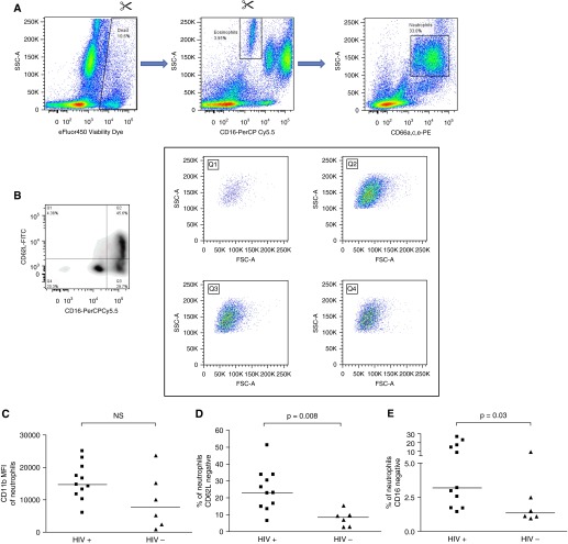
Measurements and main results: HIV infection impaired control of Mycobacterium tuberculosis by neutrophils (mean ratio of mycobacterial luminescence in neutrophil samples vs. serum controls at 1 hour in HIV-infected participants, 0.88 ± 0.13 vs. HIV-uninfected participants, 0.76 ± 0.14; P = 0.01; at 24 hours, 0.82 ± 0.13 vs. 0.71 ± 0.13; P = 0.01). The extent of impairment correlated with log[HIV viral load]. Neutrophil cell death after 24 hours' incubation with M. tuberculosis was higher in the HIV-infected cohort (85.3 ± 11.8% vs. 57.9 ± 22.4% necrotic cells; P < 0.0001). Neutrophils from HIV-infected participants demonstrated significantly more CD62L-negative cells (median, 23.0 vs. 8.5%; P = 0.008) and CD16-negative cells (3.2 vs. 1.3%, P = 0.03). Antiretroviral therapy restored mycobacterial restriction and pattern of neutrophil death toward levels seen in HIV-uninfected persons.

Conclusions: Neutrophils in antiretroviral-naive HIV-infected persons are hyperactivated, eliminate M. tuberculosis less effectively than in HIV-uninfected individuals, and progress rapidly to necrotic cell death. These factors are ameliorated by antiretroviral therapy.

Keywords: granulocyte; necrosis; tuberculosis.


Figures

Figure 1.
Figure 1.
The impact of HIV and antiretroviral therapy on neutrophil antimycobacterial activity. (AC) Twenty antiretroviral-naive HIV-infected patients were recruited and followed for 6 months of therapy (n = 19 after baseline time point, n = 18 after Month 3). Blood was taken at baseline, 1 month, 3 months, and 6 months for determination of HIV viral load (A), CD4 count (B), and percentage (C). **P < 0.01, ***P < 0.001 (Wilcoxon signed rank test versus baseline). (D) A total of 4 × 105 CD15+ granulocytes in RPMI-1640 were incubated with 10% autologous serum and M. tuberculosis-lux. Control samples did not contain neutrophils. Mycobacterial luminescence was measured for 20 seconds after 1 hour of incubation at 37°C, and the ratio of relative light units (RLU) in neutrophil samples versus serum controls is presented. Lines represent means. (E) Data from the HIV-infected individuals in D are correlated to log[HIV viral load]. R = 0.55 (95% CI, 0.13–0.80), P = 0.015. The dotted line represents a ratio of 1 (no impact of neutrophils vs. serum controls). (F) Samples were processed as in D at the indicated intervals over 6 months of antiretroviral therapy in HIV-infected individuals; data from HIV-uninfected individuals are presented for comparison. Lines represent means; dotted line indicates ratio of 1 (no impact of neutrophils vs. serum controls).
Figure 2.
Figure 2.
The impact of neutrophil depletion from blood on mycobacterial control in HIV-infected and HIV-uninfected people. (A) Blood was taken from 20 HIV-uninfected donors, 20 antiretroviral-naive HIV-infected donors, and 18 of the same HIV-infected donors after 6 months of antiretroviral therapy. A total of 450 μl of blood depleted of CD15+ cells or undepleted (in triplicate per donor per condition), diluted 1:1 with RPMI-1640, was infected with approximately 5 × 105 cfu M. tuberculosis-lux. Samples were incubated at 37°C for 96 hours before red cell lysis and measurement of mycobacterial luminescence on at least two 100-μl aliquots. Results are presented as the ratio of mycobacterial luminescence in the CD15-depleted conditions versus nondepleted controls. Solid lines represent means; the dotted line indicates a ratio of 1 (no impact of depletion). (B) Results from the CD15-depleted condition in HIV-infected donors at baseline are correlated versus log[HIV viral load]. R = −0.67 (95% CI, −0.32 to −0.86), P = 0.001. The dotted line indicates a ratio of 1 (no impact of depletion).
Figure 3.
Figure 3.
The impact of HIV and antiretroviral therapy on M. tuberculosis–associated neutrophil cell death. (A, B) A total of 4 × 105 CD15+ granulocytes in RPMI-1640 were incubated with 10% autologous serum and M. tuberculosis-lux. After 24 hours’ incubation at 37°C, samples were labeled with Annexin V-FITC, eFluor450 Viability Dye, and CD66a,c,e-PE for 15 minutes. Displayed are the percentages of total CD66a,c,e+ events (A) or “granulocytes” as defined by forward and side scatter (B) for HIV-infected and HIV-uninfected donors classed as viable (Annexin V negative, Viability Dye negative), apoptotic (Annexin V positive, Viability Dye negative), apoptotic and necrotic (Annexin V positive, Viability Dye positive), and primary necrotic (Annexin V negative, Viability Dye positive). (C, D) Samples were processed as in (A). The mean (SD) percentages of total CD66a,c,e+ events (C) or “granulocytes” (D) are displayed for HIV-infected donors at baseline and after 1, 3, or 6 months of antiretroviral therapy classed as viable, apoptotic, apoptotic and necrotic, and primary necrotic; data from HIV-uninfected donors are shown for comparison.
Figure 4.
Figure 4.
The impact of HIV on neutrophil activation markers immediately ex vivo. (A) A total of 200 μl of blood was stained immediately after phlebotomy with fluorochromes; red cells were lysed and samples fixed before acquisition within 48 hours on a BD Fortessa flow cytometer. Singlet signals (derived from Forward Scatter [FSC] area vs. FSC height, not shown) were first gated on eFluor450 Viability Dye versus Side Scatter (SSC) to exclude dead cells. Eosinophils, defined as CD16-negative with high SSC, are excluded and then neutrophils are defined as CD66a,c,e-PE–positive events with high/intermediate SSC. (B) Neutrophils thus defined are divided into quadrants on the basis of CD62L–fluorescein isothiocyanate (FITC) and CD16-PerCP Cy5.5. The contents of each quadrant are also shown on plots of FSC versus SSC, indicating that all quadrants resemble granulocytes. (C) The median fluorescence intensity of CD11b-PE Cy7 on neutrophils is plotted for 11 HIV-infected and 6 HIV-uninfected individuals. NS = not significant. (D) The percentage of neutrophils negative for CD62L is plotted for 11 HIV-infected and 6 HIV-uninfected individuals. (E) The percentage of neutrophils negative/intermediate for CD16 is plotted for 11 HIV-infected and 6 HIV-uninfected individuals. Lines in C – E represent medians, and P values are derived from Mann-Whitney tests.

References

    1. World Health Organization. Global Health Observatory (GHO) data. How many TB cases and deaths are there? [accessed 2015 Jan]. Available from: www.who.int/gho/tb/epidemic/cases_deaths/en/
    1. World Health Organization. Global Health Observatory (GHO) Data. Number of deaths due to HIV/AIDS [accessed 2015 Jan] Available from: http://www.who.int/gho/hiv/epidemic_status/deaths/en/
    1. Martineau AR, Newton SM, Wilkinson KA, Kampmann B, Hall BM, Nawroly N, Packe GE, Davidson RN, Griffiths CJ, Wilkinson RJ. Neutrophil-mediated innate immune resistance to mycobacteria. J Clin Invest. 2007;117:1988–1994. - PMC - PubMed
    1. Lowe DM, Bandara AK, Packe GE, Barker RD, Wilkinson RJ, Griffiths CJ, Martineau AR. Neutrophilia independently predicts death in tuberculosis. Eur Respir J. 2013;42:1752–1757. - PMC - PubMed
    1. Lawn SD. AIDS in Africa: the impact of coinfections on the pathogenesis of HIV-1 infection. J Infect. 2004;48:1–12. - PubMed

Publication types

MeSH terms