. 2015 Oct 1;125(10):3819-30.
doi: 10.1172/JCI79048. Epub 2015 Sep 14.

Perhexiline activates KLF14 and reduces atherosclerosis by modulating ApoA-I production

Perhexiline activates KLF14 and reduces atherosclerosis by modulating ApoA-I production

Yanhong Guo et al. J Clin Invest. .

Abstract

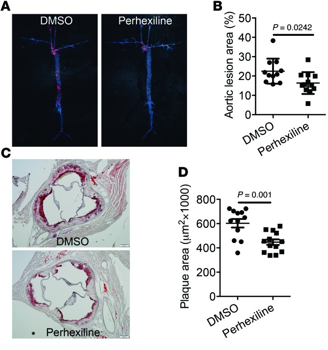
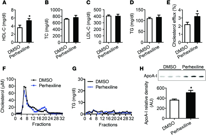
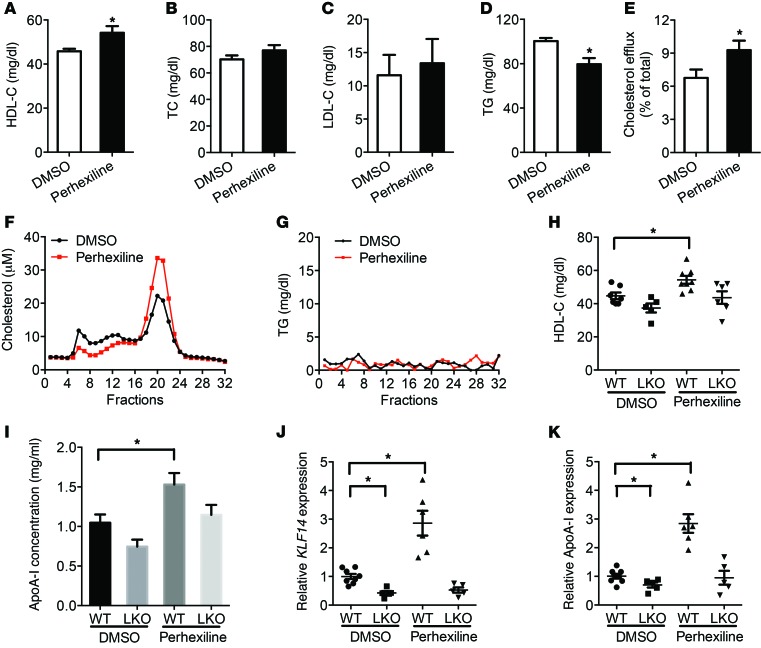
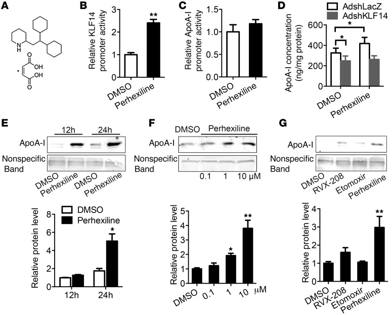
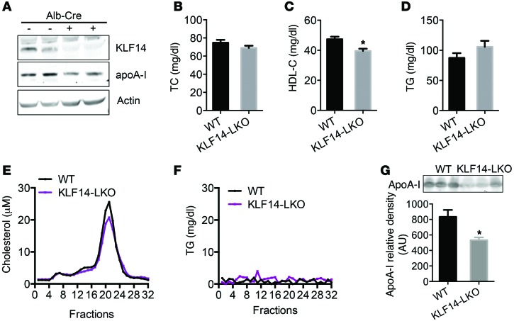
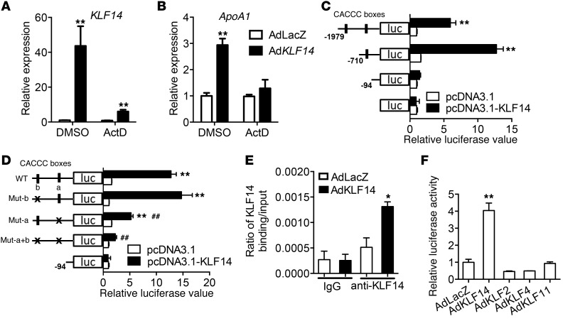
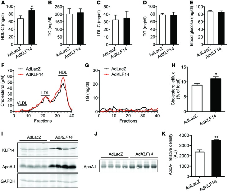
Recent genome-wide association studies have revealed that variations near the gene locus encoding the transcription factor Krüppel-like factor 14 (KLF14) are strongly associated with HDL cholesterol (HDL-C) levels, metabolic syndrome, and coronary heart disease. However, the precise mechanisms by which KLF14 regulates lipid metabolism and affects atherosclerosis remain largely unexplored. Here, we report that KLF14 is dysregulated in the liver of 2 dyslipidemia mouse models. We evaluated the effects of both KLF14 overexpression and genetic inactivation and determined that KLF14 regulates plasma HDL-C levels and cholesterol efflux capacity by modulating hepatic ApoA-I production. Hepatic-specific Klf14 deletion in mice resulted in decreased circulating HDL-C levels. In an attempt to pharmacologically target KLF14 as an experimental therapeutic approach, we identified perhexiline, an approved therapeutic small molecule presently in clinical use to treat angina and heart failure, as a KLF14 activator. Indeed, in WT mice, treatment with perhexiline increased HDL-C levels and cholesterol efflux capacity via KLF14-mediated upregulation of ApoA-I expression. Moreover, perhexiline administration reduced atherosclerotic lesion development in apolipoprotein E-deficient mice. Together, these results provide comprehensive insight into the KLF14-dependent regulation of HDL-C and subsequent atherosclerosis and indicate that interventions that target the KLF14 pathway should be further explored for the treatment of atherosclerosis.


Figures

Figure 7
Figure 7. Administration of perhexiline reduces atherosclerosis development in Apoe–/– mice.
Apoe–/– mice were placed on a HCD for 12 weeks and then were treated with DMSO or perhexiline at 10 mg/kg for 6 weeks (3 times a week) via gavage administration with continuous HCD. Perhexiline-treated mice exhibited decreased oil red O–stained lesions in the whole aorta (A) as well as reduced cross-sectional plaque area in the aortic sinus (C). Scale bars: 100 μm. Quantified en face (B) and histology (D) data are shown. Data represent mean ± SEM (n = 11–12). Student’s t test.
Figure 6
Figure 6. Administration of perhexiline increased HDL-C and ApoA-I levels and enhanced serum cholesterol efflux capacity in Apoe–/– mice.
Apoe–/– mice were placed on a HFD for 12 weeks and then were treated with DMSO or perhexiline at 10 mg/kg for 6 weeks (3 times a week) via gavage administration with continuous HFD (n = 15 per group). Plasma samples were collected and subjected individually to analytical chemistry to measure HDL-C (A), TC (B), LDL-C (C), and TG (D). *P < 0.05, Student’s t test. (E) The ABCA1-mediated cholesterol efflux capacity of serum from DMSO- or perhexiline-treated mice is expressed as the percentage of cholesterol efflux of total cell cholesterol (n = 15 per group). *P < 0.05, Student’s t test. The pooled serum from DMSO- or perhexiline-treated mice was analyzed by HPLC (fractions 1 to 32) and the cholesterol (F) and TG (G) levels in each fraction were measured. (H) Representative Western blot and quantifications of ApoA-I levels by Western blot analysis in 3 μl of plasma samples from mice treated with DMSO or perhexiline (n = 14 per group). *P < 0.05, Student’s t test. Values represent mean ± SEM.
Figure 5
Figure 5. Administration of perhexiline increased HDL-C levels in vivo.
C57BL/6J mice placed on a HFD for 12 weeks were treated with DMSO or perhexiline maleate salt (10 mg/kg/d) for 5 consecutive days by gavage administration, and plasma samples were collected at day 7 (n = 10 per group). HDL-C (A), TC (B), LDL-C (C), and TG (D) levels were measured. *P < 0.05, Student’s t test. (E) The ABCA1-mediated cholesterol efflux capacity of serum from DMSO- or perhexiline-treated mice is expressed as the percentage of cholesterol efflux of total cell cholesterol (n = 10 per group). *P < 0.05, Student’s t test. Pooled serum samples from DMSO- or perhexiline-treated mice were assayed by HPLC, and cholesterol (F) and TG (G) levels (fractions 1 to 32) were determined. (HK) KLF14-LKO and littermate control mice were treated with DMSO or perhexiline maleate salt (10 mg/Kg/d) for 5 consecutive days by gavage administration, and plasma samples were collected at day 7 (n = 5–8 for each genotype). HDL-C levels were determined (H) and ApoA-I levels were quantified by Western blot analysis (I) (n = 5–8 for each genotype). (J and K) qRT-PCR analysis showing the expression levels of Klf14 and Apoa1 in indicated groups. Data are expressed relative to 18S RNA (n = 5–8 for each genotype). Values represent mean ± SEM. *P < 0.05; **P < 0.01, 2-way ANOVA and multiple comparisons.
Figure 4
Figure 4. Drug screening identifies perhexiline as an activator of KLF14.
(A) Diagram of the chemical structure of the perhexiline maleate salt. (B and C) Luciferase activity of reporters was analyzed in HepG2 cells transfected with pGL4-KLF-luc or pGL4–ApoA-I–Luc constructs after 12 hours treatment with 10 μM perhexiline or DMSO. **P < 0.01, Student’s t test. Values represent mean ± SEM; n = 3. (D) HepG2 cells were infected with AdshLacZ or AdshKLF14 for 48 hours and then incubated with 10 μM perhexiline for 24 hours in DMEM containing 0.2% BSA. The ApoA-I concentrations in the medium were detected by ELISA. *P < 0.05, 2-way ANOVA and multiple comparisons. Values represent mean ± SEM; n = 6. (E) HepG2 cells were treated with DMSO or perhexiline at 10 μM for indicated time points in DMEM containing 0.2% BSA, and ApoA-I production was detected by Western blot. (F) HepG2 cells were treated with DMSO or perhexiline at indicated dosage for 24 hours in DMEM containing 0.2% BSA, and ApoA-I production was detected by Western blot. (G) HepG2 cells were treated with DMSO, perhexiline, RVX-208, or etomoxir at 10 μM for 24 hours in DMEM containing 0.2% BSA, and ApoA-I production was detected by Western blot. Quantifications from 3 independent experiments are shown in EG, and values represent mean ± SEM. *P < 0.05; **P < 0.01, 2-way ANOVA and multiple comparisons.
Figure 3
Figure 3. Liver-specific deletion of Klf14 showed decreased HDL-C levels.
(A) Western blot of KLF14 and ApoA-I expression in the livers of KLF14-LKO and littermate control (WT) mice. TC (B), HDL-C (C), and TG (D) levels were determined in KLF14-LKO and littermate control mice fasted overnight (n = 5–7 for each genotype). *P < 0.05, Student’s t test. (E and F) Pooled serum samples from KLF14-LKO and WT mice were assayed by HPLC, and cholesterol and TG levels (fractions 1 to 32) were determined. (G) Representative Western blot and quantifications of ApoA-I levels by Western blot analysis in 1 μl of serum samples from WT and KLF14-LKO mice. Values represent mean ± SEM (5–7 for each genotype). *P < 0.05, Student’s t test.
Figure 2
Figure 2. KLF14 is a regulator of APOA1 expression.
HepG2 cells were infected with AdLacZ or AdKLF14 for 24 hours and then incubated in medium containing ActD (5 μg/ml) or DMSO for another 24 hours (n = 3). KLF14 (A) and APOA1 (B) mRNA levels were determined by real-time qPCR. **P < 0.01, 2-way ANOVA and multiple comparisons. (C) The structure of human APOA1 promoter used in the luciferase assays indicating 2 putative CACCC-box KLF-binding sites. Expression of KLF14 with human APOA1 promoter assay demonstrated that KLF14 significantly increased ApoA-I luciferase activity (n = 3). **P < 0.01, compared with control vector, 2-way ANOVA and multiple comparisons. (D) Mutations of the 2 putative KLF-binding sites demonstrated ApoA-I expression is dependent on KLF14 and CACCC-box binding sites (n = 3). **P < 0.01, compared with pGL4-basic vector; ##P < 0.01, compared with APOA1 promoter WT, 2-way ANOVA and multiple comparisons. (E) ChIP assay revealed significant enrichment of KLF14 protein on the human APOA1 promoter in HepG2 cells (n = 3). *P < 0.05, 2-way ANOVA and multiple comparisons. (F) Luciferase activity assay demonstrated that KLF14, not KLF2, KLF4, or KLF11, led to an increase in APOA1 promoter activity in HepG2 cells (n = 3). **P < 0.01, 2-way ANOVA and multiple comparisons. Representative of at least 3 experiments.
Figure 1
Figure 1. Overexpression of KLF14 increases both HDL-C and ApoA-I levels and cholesterol efflux capacity.
Adenoviral vectors containing LacZ (AdLacZ) or human KLF14 (AdKLF14) (5 × 10⁸ pfu per mouse) were administered via tail vein injection to C57BL/6 mice fed HFD for 12 weeks (n = 10 per group). Serum samples were collected at day 6 and subjected individually to analytical chemistry to measure HDL-C (A), TC (B), LDL-C (C), TG (D), and fasting blood glucose (E) or to determine cholesterol and TG levels from pooled samples by FPLC (fractions 1 to 40) (F and G). *P < 0.05, Student’s t test. (H) The ABCA1-mediated cholesterol efflux capacity of serum from AdKLF14- or AdLacZ-treated mice is expressed as the percentage of cholesterol efflux of total cell cholesterol (n = 10 per group). *P < 0.05, Student’s t test. Representative Western blot results show that AdKLF14-treated mice exhibited increased expression of ApoA-I levels in the liver (I) and serum (J). (K) Quantifications of ApoA-I levels in the serum from AdLacZ and AdKLF14-treated mice by Western blot (n = 10 per group). Values represent mean ± SEM. **P < 0.01, Student’s t test.

Similar articles

Cited by

References

    1. Gordon T, Castelli WP, Hjortland MC, Kannel WB, Dawber TR. High density lipoprotein as a protective factor against coronary heart disease. The Framingham Study. Am J Med. 1977;62(5):707–714. doi: 10.1016/0002-9343(77)90874-9. - DOI - PubMed
    1. Gordon DJ, et al. Bangdiwala S, and Tyroler HA. High-density lipoprotein cholesterol and cardiovascular disease. Four prospective American studies. Circulation. 1989;79(1):8–15. doi: 10.1161/01.CIR.79.1.8. - DOI - PubMed
    1. McQueen MJ, et al. Lipids, lipoproteins, and apolipoproteins as risk markers of myocardial infarction in 52 countries (the INTERHEART study): a case-control study. Lancet. 2008;372(9634):224–233. doi: 10.1016/S0140-6736(08)61076-4. - DOI - PubMed
    1. Yusuf S, et al. Effect of potentially modifiable risk factors associated with myocardial infarction in 52 countries (the INTERHEART study): case-control study. Lancet. 2004;364(9438):937–952. doi: 10.1016/S0140-6736(04)17018-9. - DOI - PubMed
    1. Boekholdt SM, et al. Levels and changes of HDL cholesterol and apolipoprotein A-I in relation to risk of cardiovascular events among statin-treated patients: a meta-analysis. Circulation. 2013;128(14):1504–1512. doi: 10.1161/CIRCULATIONAHA.113.002670. - DOI - PMC - PubMed

Publication types

MeSH terms