Comparative Study
. 2015 Oct 1;125(10):3928-40.
doi: 10.1172/JCI79271. Epub 2015 Sep 14.

Inhibition of hyaluronan synthesis restores immune tolerance during autoimmune insulitis

Comparative Study

Inhibition of hyaluronan synthesis restores immune tolerance during autoimmune insulitis

Nadine Nagy et al. J Clin Invest. .

Abstract

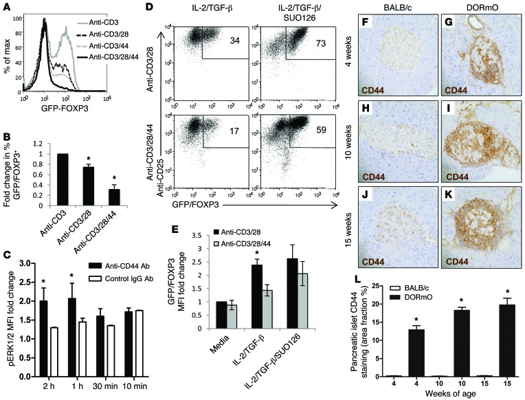
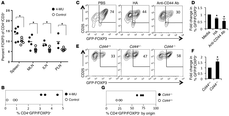
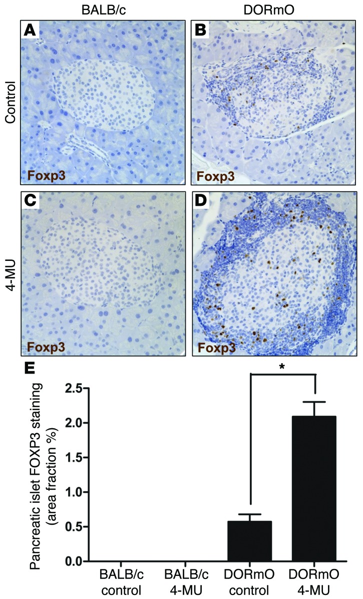
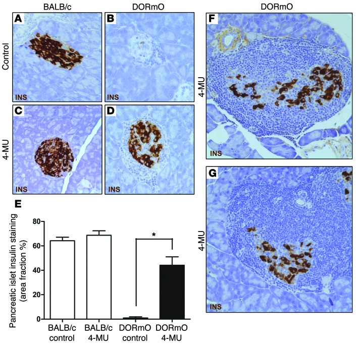
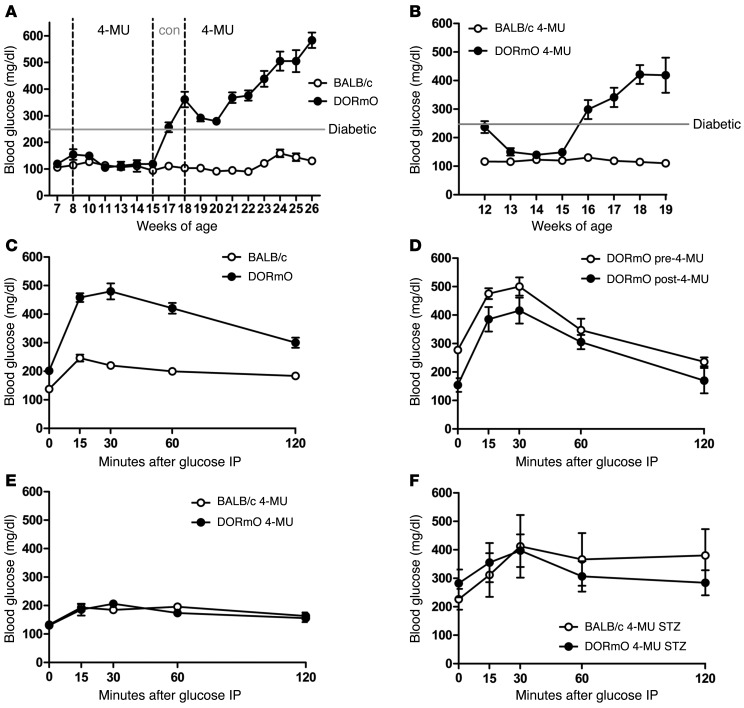
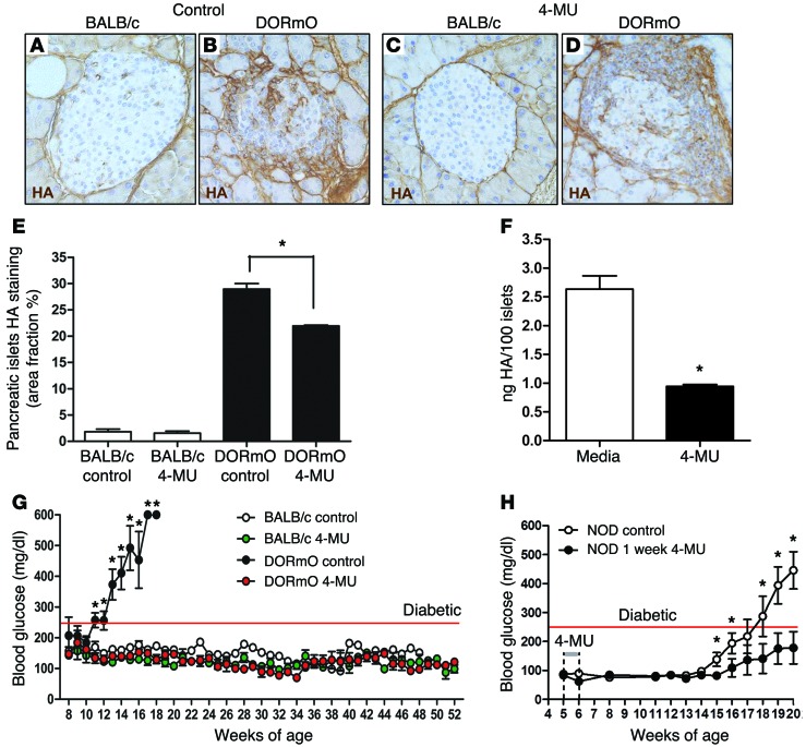
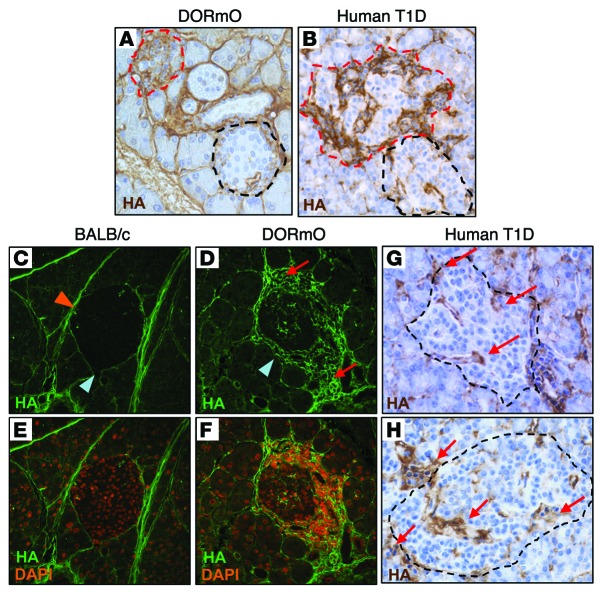
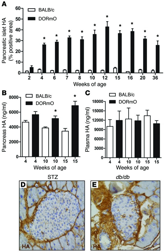
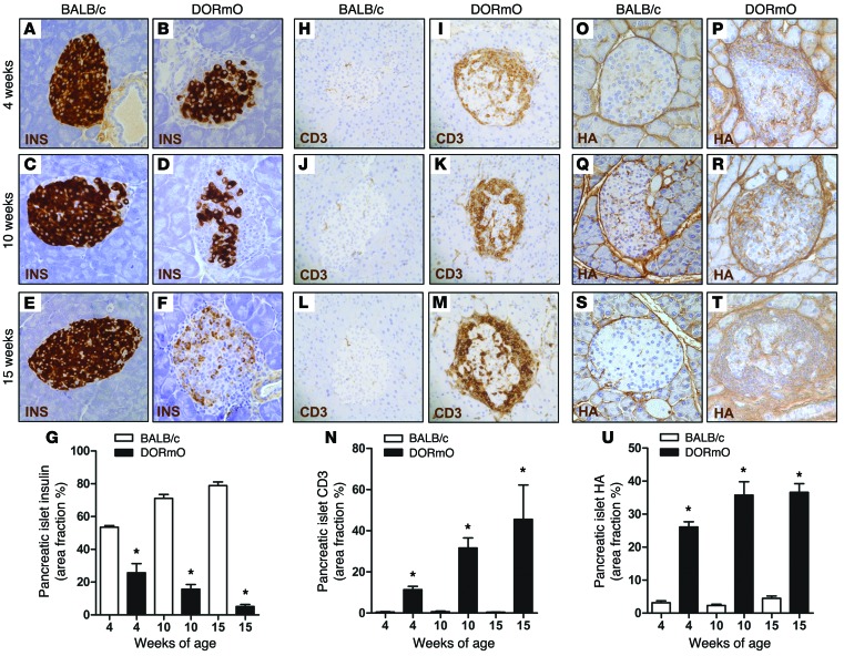
We recently reported that abundant deposits of the extracellular matrix polysaccharide hyaluronan (HA) are characteristic of autoimmune insulitis in patients with type 1 diabetes (T1D), but the relevance of these deposits to disease was unclear. Here, we have demonstrated that HA is critical for the pathogenesis of autoimmune diabetes. Using the DO11.10xRIPmOVA mouse model of T1D, we determined that HA deposits are temporally and anatomically associated with the development of insulitis. Moreover, treatment with an inhibitor of HA synthesis, 4-methylumbelliferone (4-MU), halted progression to diabetes even after the onset of insulitis. Similar effects were seen in the NOD mouse model, and in these mice, 1 week of treatment was sufficient to prevent subsequent diabetes. 4-MU reduced HA accumulation, constrained effector T cells to nondestructive insulitis, and increased numbers of intraislet FOXP3+ Tregs. Consistent with the observed effects of 4-MU treatment, Treg differentiation was inhibited by HA and anti-CD44 antibodies and rescued by 4-MU in an ERK1/2-dependent manner. These data may explain how peripheral immune tolerance is impaired in tissues under autoimmune attack, including islets in T1D. We propose that 4-MU, already an approved drug used to treat biliary spasm, could be repurposed to prevent, and possibly treat, T1D in at-risk individuals.


Figures

Figure 9
Figure 9. 4-MU treatment promotes FOXP3 induction.
(A) FOXP3 levels following induction from CD4+GFP/FOXP3 T cell precursors performed in the setting of anti-CD3, with or without anti-CD28 and/or anti-CD44 antibody costimulation. (B) Pooled data for 4 independent experimental replicates for the representative data in A. (C) Fold change in pERK1/2 MFI over time following CD44 crosslinking. The data shown incorporate 3 experimental replicates. (D) CD25 and GFP/FOXP3 levels following activation of CD4+GFP/FOXP3 T cells in the setting of TGF-β and IL-2, with or without CD44 costimulation and/or the ERK1/2 inhibitor SUO126. (E) Pooled data for 6 independent experimental replicates for the representative data in D. (FK) CD44 staining of representative pancreatic tissue sections from BALB/c (control) or DORmO mice fed either 4-MU or control chow. (L) Average CD44+ area of islets for these mice. At least 25 islets were visualized per mouse (n = 6). Original magnification, ×40. Data represent mean ± SEM; *P < 0.05 vs. respective control and control for each time point by unpaired t test.
Figure 8
Figure 8. 4-MU treatment relieves CD44-mediated inhibition of FOXP3 induction.
(A) Percentage of GFP/FOXP3+ Tregs of total CD4+ T cells in BALB/c mice fed 4-MU or control chow for 2 weeks (n = 5–6 mice per group). (B) In vivo induction of FOXP3+ Tregs assessed 4 days after transfer of GFP/FOXP3CD4+ T cells into Rag–/– hosts given 4-MU or control chow (n = 3 Rag–/– recipient animals). Data are from the spleens of recipient animals. (C) CD25 and FOXP3 expression by CD4+GFP/FOXP3 T cells activated for 72 hours with or without plate-bound HA or anti-CD44 antibody. (D) Pooled data for 3 independent experimental replicates for the representative data in C. (E) FOXP3 induction using Cd44+/+, Cd44–/+, or Cd44–/– precursors. (F) Pooled data for 3 independent experimental replicates for the representative data in E. (G) In vivo induction of FOXP3 assessed using cotransfer of equivalent numbers of GFP/FOXP3CD4+Cd44+/+CD45.1 and GFP/FOXP3CD4+Cd44–/–CD45.2 T cells into Rag–/– hosts. After 4 days, the numbers of induced CD3+GFP/FOXP3+ Tregs in the spleens of recipient animals were assessed and the ratio of Cd44–/– Tregs versus Cd44+/+ Tregs was determined (n = 3 Rag–/– recipient animals). Data represent mean ± SEM; *P < 0.05 vs. respective control by unpaired t test.
Figure 7
Figure 7. 4-MU treatment increases islet FOXP3+ Tregs.
(AD) FOXP3 staining of representative pancreatic tissue sections from DORmO and BALB/c mice fed either 4-MU or control chow. (E) Average FOXP3+ islet area of these mice. At least 25 islets were visualized per mouse, and data are for 6 mice per condition. Original magnification, ×40. Data represent mean ± SEM; *P < 0.05 vs. respective control by unpaired t test.
Figure 6
Figure 6. 4-MU treatment promotes nondestructive insulitis.
(AD) Insulin staining of representative pancreatic tissue sections from DORmO and BALB/c mice fed either 4-MU or control chow. (E) Average insulin+ area of islets for these mice. 25 islets were visualized per mouse, and staining and data are for 6 mice per condition. (F and G) Representative images of insulin staining of pancreatic islets from 15-week-old DORmO mice treated with 4-MU for 7 weeks. Original magnification, ×40. Data represent mean ± SEM; *P < 0.05 vs. respective control by unpaired t test.
Figure 5
Figure 5. 4-MU treatment prevents progression of insulitis but does not cure established diabetes.
(A) Blood glucose of DORmO and control mice started on 4-MU at 8 weeks of age, taken off 4-MU between 15 and 18 weeks of age, and restarted on 4-MU thereafter (n = 10). (B) Blood glucose of DORmO and BALB/c (control) mice fed 4-MU chow, beginning at 12 weeks of age (n = 10). (C) Blood glucose following IPGTT for control chow–fed BALB/c and DORmO mice at 10 weeks of age (n = 8 mice per group). (D) Blood glucose following IPGTT of DORmO mice from C treated for 2 weeks with 4-MU or control chow. (E) Blood glucose following IPGTT of BALB/c and DORmO mice at 10 weeks of age following 4-MU treatment for 2 weeks (n = 6). (F) Blood glucose following IPGTT for the same mice as in E, now made hyperglycemic by STZ treatment (n = 6). Data represent mean ± SEM.
Figure 4
Figure 4. Inhibition of HA synthesis prevents progression to autoimmune diabetes.
(AD) Representative HA staining of pancreatic tissue from BALB/c (control) and DORmO mice fed 4-MU chow or control chow, beginning at 8 weeks of age, a month after the typical onset of insulitis in this model. (E) Average islet HA+ area in these same mice. 25 islets were visualized per mouse, and staining and data are for 6 mice per condition. (F) HA content in islets from BALB/c mice cultured with 50 μg/ml 4-MU or media alone. Data are for 100 islets, and experiments were done in triplicate. (G) Blood glucose of DORmO and BALB/c (control) mice fed 4-MU chow or control chow, beginning at 8 weeks of age, and maintained on 4-MU for 1 year (n = 12 mice per group). (H) Blood glucose of NOD mice fed with control chow or fed with control chow including a 1 week of 4-MU treatment from 5 to 6 weeks of age (n = 10 mice per group). Original magnification, ×40. Data represent mean ± SEM; *P < 0.05 vs. respective control and control for each time point by unpaired t test.
Figure 3
Figure 3. HA deposits characterize sites of insulitis in DORmO and human T1D.
(A and B) HA staining in pancreatic tissue isolated from (A) a DORmO mouse and (B) a human cadaveric donor with T1D. Infiltrated islets are circled in red; unaffected islets are circled in black. (C and D) HA staining of representative BALB/c and DORmO islets, demonstrating interstitial (orange arrowhead) and peri-islet (blue arrowheads) patterns of HA distribution. HA associated with lymphocytic infiltrates (red arrows) was only seen in DORmO mice. (E and F) Costaining of HA and DAPI, demonstrating HA accumulation in association with insulitis. (G and H) HA deposits in human insulitis (red arrows) from two cadaveric donors with T1D. Original magnification, ×40.
Figure 2
Figure 2. Islet HA deposition is temporally associated with insulitis and not hyperglycemia.
(A) Percentage of islet area positive for HA staining of DORmO and BALB/c (control) mice over time (n = 6–15). (B) Pancreas and (C) plasma HA content in BALB/c and DORmO mice over time (n = 6). (D and E) Representative HA staining of pancreatic islet tissue from (D) a BALB/c mouse 1 week after STZ treatment and (E) a 12-week-old diabetic db/db mouse. Original magnification, ×40. Data represent mean ± SEM; *P < 0.05 vs. control for each time point by unpaired t test.
Figure 1
Figure 1. Islet HA accumulates in tandem with progressive autoimmune insulitis in DORmO mice.
Representative histologic staining of pancreatic tissue from BALB/c (control) and DORmO mice and average islet area positive over time for (AG) insulin (INS), (HN) CD3, and (OU) HA. For G, N, and U, at least 25 islets were visualized per mouse, and staining and data are from n = 6–8 mice per condition. Original magnification, ×40. Data represent mean ± SEM; *P < 0.05 vs. control for each time point by unpaired t test.

References

    1. Wildin RS, et al. X-linked neonatal diabetes mellitus, enteropathy and endocrinopathy syndrome is the human equivalent of mouse scurfy. Nat Genet. 2001;27(1):18–20. doi: 10.1038/83707. - DOI - PubMed
    1. Ziegler AG, Nepom GT. Prediction and pathogenesis in type 1 diabetes. Immunity. 2010;32(4):468–478. doi: 10.1016/j.immuni.2010.03.018. - DOI - PMC - PubMed
    1. Putnam AL, et al. Expansion of human regulatory T-cells from patients with type 1 diabetes. Diabetes. 2009;58(3):652–662. doi: 10.2337/db08-1168. - DOI - PMC - PubMed
    1. Glisic S, Ehlenbach S, Jailwala P, Waukau J, Jana S, Ghosh S. Inducible regulatory T cells (iTregs) from recent-onset type 1 diabetes subjects show increased in vitro suppression and higher ITCH levels compared with controls. Cell Tissue Res. 2010;339(3):585–595. doi: 10.1007/s00441-009-0900-0. - DOI - PubMed
    1. Zhou X, et al. Instability of the transcription factor Foxp3 leads to the generation of pathogenic memory T cells in vivo. Nat Immunol. 2009;10(9):1000–1007. doi: 10.1038/ni.1774. - DOI - PMC - PubMed

Publication types

MeSH terms