. 2015 Sep 14;10(9):e0137711.
doi: 10.1371/journal.pone.0137711. eCollection 2015.

Identification of Tetraazacyclic Compounds as Novel Potent Inhibitors Antagonizing RORγt Activity and Suppressing Th17 Cell Differentiation

Affiliations

Identification of Tetraazacyclic Compounds as Novel Potent Inhibitors Antagonizing RORγt Activity and Suppressing Th17 Cell Differentiation

Qingfeng Ding et al. PLoS One. .

Abstract

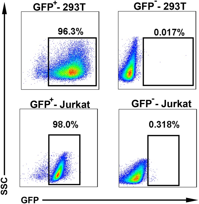
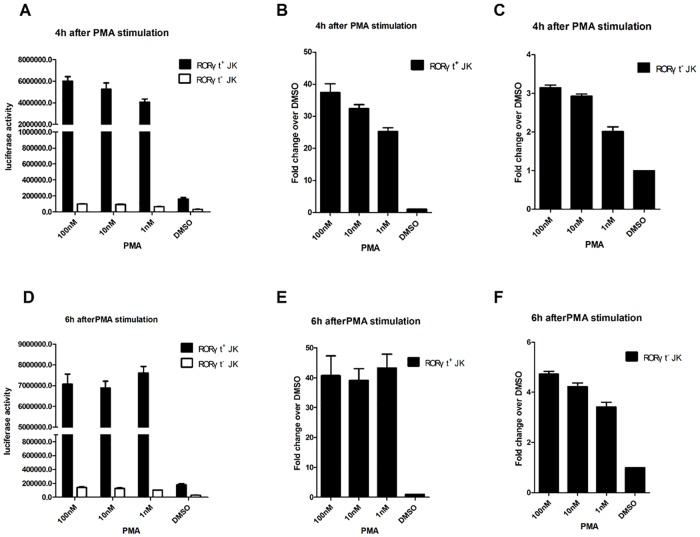
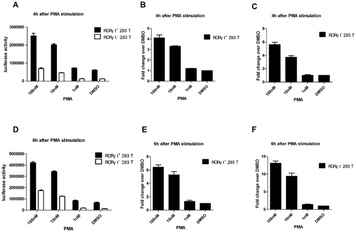
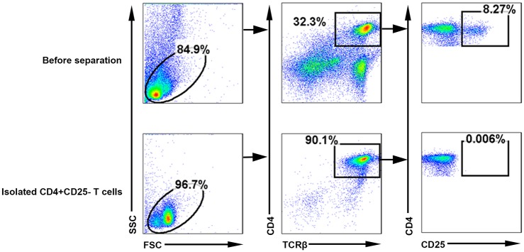
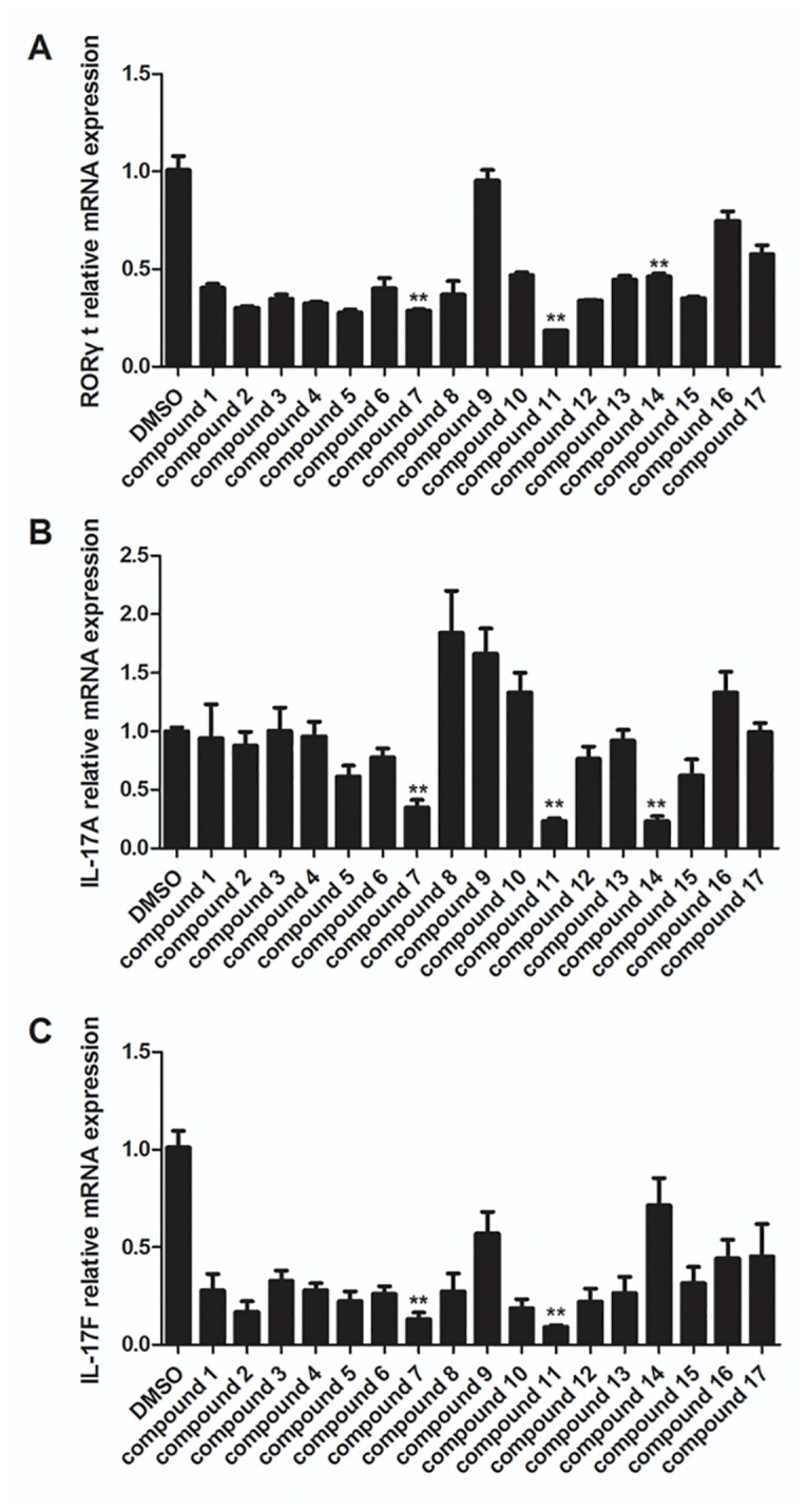
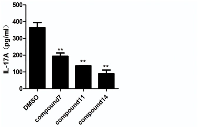
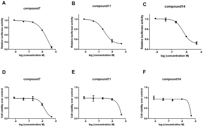
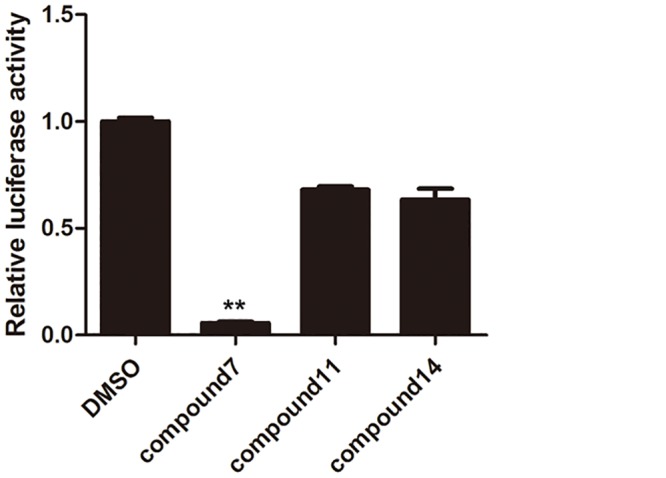
CD4+ T-helper cells that produce interleukin-17 (Th17 cells) are characterized as pathological T-helper cells in autoimmune diseases. Differentiation of human and mouse Th17 cells requires a key transcription regulator, retinoic acid receptor-related orphan receptor γt (RORγt), which is a potential therapeutic target for autoimmune diseases. To develop a therapeutic agent for Th17-mediated autoimmune diseases, we have established a high-throughput screening (HTS) assay for candidate screening, in which the luciferase activity in RORγt-LBD positive and negative Jurkat cells were analyzed to evaluate induction of RORγt activity by compounds. This technique was applied to screen a commercially-available drug-like chemical compound library (Enamine) which contains 20155 compounds. The screening identified 17 compounds that can inhibit RORγt function in the HTS screen system. Of these, three tetraazacyclic compounds can potently inhibit RORγt activity, and suppress Th17 differentiation and IL-17 production. These three candidate compounds could significantly attenuate the expression of the Il17a by 65%- 90%, and inhibit IL-17A secretion by 47%, 63%, and 74%, respectively. These compounds also exhibited a potent anti-RORγt activity, with EC50 values of 0.25 μM, 0.67 μM and 2.6 μM, respectively. Our data demonstrated the feasibility of targeting the RORγt to inhibit Th17 cell differentiation and function with these tetraazacyclic compounds, and the potential to improve the structure of these compounds for autoimmune diseases therapeutics.


Conflict of interest statement

Competing Interests: The authors have declared that no competing interests exist.

Figures

Fig 1
Fig 1. Establishment of the 293T-RORγt and Jurkat-RORγt reporter cell lines.
The pGL4.31 reporter plasmids were transfected into 293T cells. The transfected cells were then selected in medium containing hygromycin B to obtain a stable clone which express pGL4.31 reporter gene. Then pBIND-RORγtLBD-IRES-GFP plasmids were also transfected into the stable clone. The second transfected cells were sorted by GFP. GFP+-293T stable cell purity was determined >96% by flow cytometry analysis, which yielded a RORγt+-293T (RORγt-LBD positive 293T cells) stable reporter cell line, and GFP--293T stable cells were also sorted and used as a RORγt negative control cell line (Fig 1 top panel). Similar to this procedure, Jurkat cells were also transfected with pGL4.31 reporter plasmids by electroporation and selected in RPMI 1640 medium containing hygromycin B. And then the PBIND-RORγtLBD-IRES-GFP plasmids were transfected into the stable Jurkat clone and were sorted by GFP. GFP+-Jurkat stable cell purity was determined >96% by flow cytometry analysis and GFP--Jurkat stable cells were also sorted (Fig 1 bottom panel).
Fig 2
Fig 2. Validation of the Jurkat-RORγt-LBD stable cell lines.
RORγt+-Jurkat (RORγt-LBD positive Jurkat cells) and RORγt--Jurkat (RORγt-LBD negative Jurkat cells) stable cells (1x10⁵) were seeded onto 12-well plates and incubated in 1 mL RPMI 1640 complete medium overnight. The cells were cultured with different concentrations of PMA (100 nM, 10 nM, 1 nM) or DMSO (vehicle control) for 4hr. The total protein was extracted to determine luciferase activity (A). The fold changes in luciferase activity compared to the DMSO control were determined in RORγt+-Jurkat cells and RORγt--Jurkat cells, when the cells were cultured with different concentrations of PMA (100 nM, 10 nM, 1 nM) or DMSO for 4hr (B, C). RORγt+ and RORγt--Jurkat cells (1 × 10⁵) were seeded onto 12-well plates and incubated in 1 mL RPMI 1640 complete medium overnight. Then the cells were cultured with different concentrations of PMA (100 nM, 10 nM, 1 nM) or DMSO (vehicle control) for 6 hr, at which point total protein was extracted to determine luciferase activity (D). The fold changes in luciferase activity upon exposure to compounds vs DMSO were determined in RORγt+-Jurkat cells and RORγt--Jurkat cells using different concentrations of PMA (100 nM, 10 nM, 1 nM) or DMSO (vehicle control) for 6hr (E, F). The results are shown as means ± SEM.
Fig 3
Fig 3. Validation of the 293T-RORγt-LBD stable cell lines.
RORγt+-293T (RORγt-LBD positive 293T cells) and RORγt--293T (RORγt-LBD negative 293T cells) stable cells (1x10⁵) were seeded onto 12-well plates and incubated in 1 mL DMEM complete medium overnight. Then the cells were cultured with different concentrations of PMA (100 nM, 10 nM, 1 nM) or DMSO for 4h. The total cell proteins were extracted and luciferase activity was determined (A). The fold changes in luciferase activity compared to the DMSO control were determined when the RORγt+-293T and RORγt--293T cells were cultured with different concentrations of PMA (100 nM, 10 nM, 1 nM) or DMSO (vehicle control) for 4h (B, C). RORγt+-293T and RORγt--293T stable cells (1x10⁵) were seeded onto 12-well plates as above. Then the cells were cultured with different concentrations of PMA (100 nM, 10 nM, 1 nM) or DMSO (vehicle control) for 6h. Total cell proteins were extracted and luciferase activity was determined (D). The fold changes in luciferase activity upon exposure to compounds vs DMSO were determined in RORγt+-293T and RORγt--293Tcells using different concentrations of PMA (100 nM, 10 nM, 1 nM) or DMSO (vehicle control) for 6 h (E, F). The results are shown as mean ± SEM.
Fig 4
Fig 4. Purification of the CD4+CD25- T cells.
Single-cell suspensions from spleens of 12-week-old C57BL/6 were made by crushing the spleen through a cell strainer, and red blood cells (RBCs) were lysed with an RBC lysis buffer. CD4+ T cells were then purified using MACS magnetic cell column with a CD4+ T cell isolation kit. And about 90% cells were CD4+ CD25- T cell after isolation. The proportion was significantly higher than before separation, which was only about 32%.
Fig 5
Fig 5. Three candidate compounds inhibit mouse Th17 cell differentiation.
CD4+CD25- T cells were isolated from spleens of 8–12 week old mice using MACS magnetic cell column with a CD4+ T cell isolation kit. CD4+CD25- T cells were cultured under Th17 polarizing conditions with vehicle control and hit compounds (5 μM) as described in the methods section. RORγt (A), IL-17A (B) and IL-17F (C) expression was quantified and normalized to GAPDH. The results are shown as mean ± SEM; ** P< 0.01.
Fig 6
Fig 6. Three tetraazacyclic compounds inhibit IL-17A secretion.
CD4+ T cells were cultured under Th17 polarizing conditions and the effect on IL-17A secretion in the supernatant was assessed by enzyme-linked immunosorbent assay (ELISA). The concentrations of IL-17A secreted from Th17 cells in presence of tetraazacyclic compounds at 5 μm or DMSO were determined. The results are shown as mean ± SEM; ** P< 0.01.
Fig 7
Fig 7. EC50 and CC50 of novel tetraazacyclic compounds.
RORγt+-Jurkat cells were seeded onto 96-well round-bottom plates (2x10⁴) overnight and incubated with the compounds titrated at 5-fold gradient final concentrations of 5 μM to 8 nM. 6 h later, relative luciferase activity was recorded to obtain the EC50 values of compounds 7, 11 and 14 (A, B, C, respectively). Jurkat cells (wild type) were seeded onto 96-well round-bottom plates (2x10⁴) overnight and incubated with the compounds at 5-fold gradient concentrations. 48 h treatment later, MTT was added onto 96-well plate and incubated at 37°C for 4 h. MTT was cleaved by live cells to a colored formazan product. After centrifugation (1500rpm, 5min), the supernatant was discarded. 100 μl DMSO was added to dissolve the formazan product and the solution was gently shaken for 10 min. Absorbance at 570 nm wavelength was recorded to identify CC50 values of compounds 7, 11 and 14 (D, E, F, respectively). The results are shown as mean ± SEM.
Fig 8
Fig 8. Inhibition of RORγt activity in 293T cells.
RORγt+-293T stable cells were culture in the presence of hit compounds (5 μM) and DMSO (vehicle control) for 6 h. Then the luciferase activity in RORγt+-293T cells were recorded. The results can be used as an evaluation marker to denote T cell-specific effects of these tetraazacyclic compounds. The results are shown as mean ± SEM; ** P< 0.001.

References

    1. Jetten AM, Kurebayashi S, Ueda E. The ROR nuclear orphan receptor subfamily: critical regulators of multiple biological processes. Progress in nucleic acid research and molecular biology. 2001;69:205–47. Epub 2001/09/12. . - PubMed
    1. Stockinger B, Veldhoen M. Differentiation and function of Th17 T cells. Current opinion in immunology. 2007;19(3):281–6. Epub 2007/04/17. 10.1016/j.coi.2007.04.005 . - DOI - PubMed
    1. McGeachy MJ, Cua DJ. Th17 cell differentiation: the long and winding road. Immunity. 2008;28(4):445–53. Epub 2008/04/11. 10.1016/j.immuni.2008.03.001 . - DOI - PubMed
    1. Leppkes M, Becker C, Ivanov II, Hirth S, Wirtz S, Neufert C, et al. RORgamma-expressing Th17 cells induce murine chronic intestinal inflammation via redundant effects of IL-17A and IL-17F. Gastroenterology. 2009;136(1):257–67. Epub 2008/11/11. 10.1053/j.gastro.2008.10.018 . - DOI - PubMed
    1. Li Y, Jiang L, Zhang S, Yin L, Ma L, He D, et al. Methotrexate attenuates the Th17/IL-17 levels in peripheral blood mononuclear cells from healthy individuals and RA patients. Rheumatology international. 2012;32(8):2415–22. Epub 2011/06/22. 10.1007/s00296-011-1867-1 . - DOI - PubMed

Publication types

MeSH terms

Substances