. 2015 Sep 14;10(9):e0137894.
doi: 10.1371/journal.pone.0137894. eCollection 2015.

Retinoic Acid Activity in Undifferentiated Neural Progenitors Is Sufficient to Fulfill Its Role in Restricting Fgf8 Expression for Somitogenesis

Affiliations

Retinoic Acid Activity in Undifferentiated Neural Progenitors Is Sufficient to Fulfill Its Role in Restricting Fgf8 Expression for Somitogenesis

Thomas J Cunningham et al. PLoS One. .

Abstract

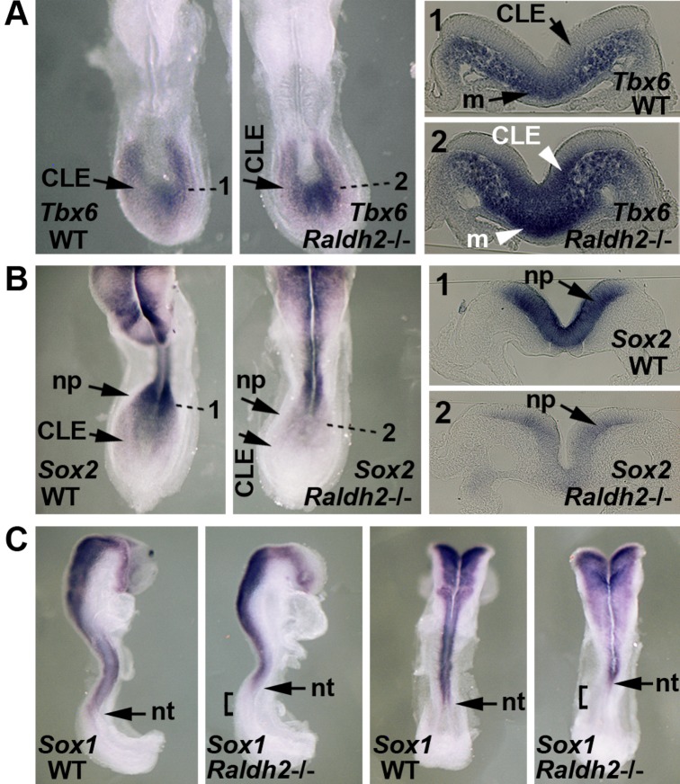
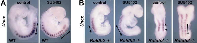
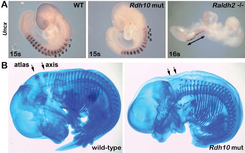
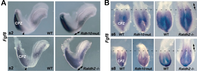
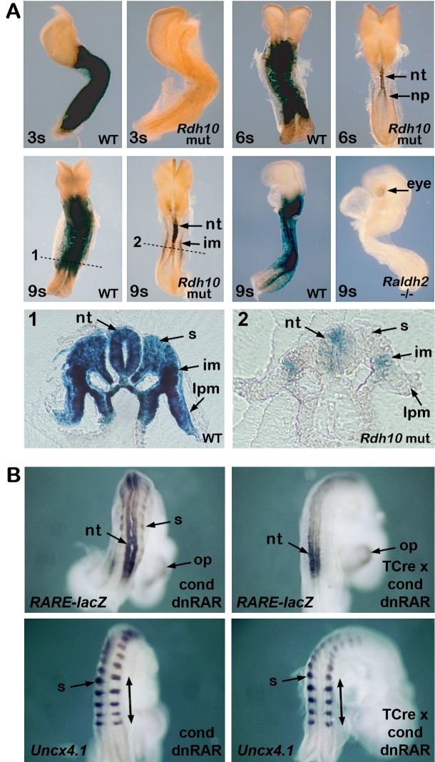
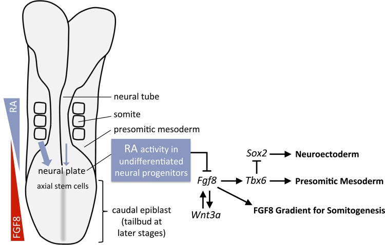
Bipotent axial stem cells residing in the caudal epiblast during late gastrulation generate neuroectodermal and presomitic mesodermal progeny that coordinate somitogenesis with neural tube formation, but the mechanism that controls these two fates is not fully understood. Retinoic acid (RA) restricts the anterior extent of caudal fibroblast growth factor 8 (Fgf8) expression in both mesoderm and neural plate to control somitogenesis and neurogenesis, however it remains unclear where RA acts to control the spatial expression of caudal Fgf8. Here, we found that mouse Raldh2-/- embryos, lacking RA synthesis and displaying a consistent small somite defect, exhibited abnormal expression of key markers of axial stem cell progeny, with decreased Sox2+ and Sox1+ neuroectodermal progeny and increased Tbx6+ presomitic mesodermal progeny. The Raldh2-/- small somite defect was rescued by treatment with an FGF receptor antagonist. Rdh10 mutants, with a less severe RA synthesis defect, were found to exhibit a small somite defect and anterior expansion of caudal Fgf8 expression only for somites 1-6, with normal somite size and Fgf8 expression thereafter. Rdh10 mutants were found to lack RA activity during the early phase when somites are small, but at the 6-somite stage RA activity was detected in neural plate although not in presomitic mesoderm. Expression of a dominant-negative RA receptor in mesoderm eliminated RA activity in presomitic mesoderm but did not affect somitogenesis. Thus, RA activity in the neural plate is sufficient to prevent anterior expansion of caudal Fgf8 expression associated with a small somite defect. Our studies provide evidence that RA restriction of Fgf8 expression in undifferentiated neural progenitors stimulates neurogenesis while also restricting the anterior extent of the mesodermal Fgf8 mRNA gradient that controls somite size, providing new insight into the mechanism that coordinates somitogenesis with neurogenesis.


Conflict of interest statement

Competing Interests: The authors have declared that no competing interests exist.

Figures

Fig 1
Fig 1. Loss of RA disrupts expression of axial stem cell niche markers.
Shown is a comparison of E8.25 (5-somite) wild-type (WT) and Raldh2-/- embryos. (A) Tbx6 mRNA; dotted lines indicate transverse sections through whole-mount stained embryos showing that loss of RA results in the appearance of ectopic Tbx6 expression in the caudal lateral epiblast (CLE) and expansion of Tbx6+ presomitic mesoderm along the dorsoventral axis. (B) Sox2 mRNA; dotted lines indicate transverse sections showing that loss of RA down-regulates Sox2 expression in neural plate (np); also, down-regulation in CLE is observed in whole-mount. (C) Sox1 mRNA; the bar shows that loss of RA results in loss of Sox1 expression in posterior neural tube (nt) adjoining the neural plate.
Fig 2
Fig 2. RA antagonism of caudal FGF signaling rescues somitogenesis defect.
(A-B) Wild-type (WT) and Raldh2-/- embryos at the 6–9 somite stages were cultured for 12 h in the presence of SU5402 (20 micromolar) or DMSO vehicle control, then processed to visualize mRNA for Uncx gene expressed in the posterior domain of each somite [26,27] to monitor somite length along the anteroposterior axis. Somite length was compared by measuring the bars bracketing the caudal-most 5 somites generated in culture.
Fig 3
Fig 3. Rdh10 mutants exhibit a small somite defect during early but not late stages.
(A) Uncx mRNA in WT, Rdh10 mutants, and Raldh2-/- embryos at the 15–16 somite stage. Numbers marking the first 6 somites of WT and Rdh10 mutants reveal a temporary shortening of somite size along the anteroposterior axis in the Rdh10 mutant; arrows mark the region displaying a much larger region of small somites in Raldh2-/- embryos. (B) Alcian blue staining of E14.5 wild-type and Rdh10 mutant embryos was performed; arrows indicate that the mutant lacks the atlas and axis vertebrae derived from somites 5 and 6, respectively.
Fig 4
Fig 4. Rdh10 mutants exhibit ectopic caudal Fgf8 expression at early but not late stages.
(A) Fgf8 mRNA in 2-somite (2s) embryos. In WT, arrowheads mark the normal anterior border for caudal Fgf8 expression. In mutants, arrows mark regions of expanded Fgf8 expression within and anterior to its normal caudal domain. (B) Fgf8 mRNA in the caudal region (anterior side up) at stages somite-6 (s6) and somite-8 (s8). Dotted lines mark the normal anterior border for caudal Fgf8 expression in WT. Arrows mark regions of expanded Fgf8 expression anterior to its normal caudal domain seen for Raldh2-/- embryos but not Rdh10 mutants. CPZ, caudal progenitor zone.
Fig 5
Fig 5. RA signaling in neural plate is sufficient to control somitogenesis.
(A) RA activity was visualized in embryos carrying the RARE-lacZ RA-reporter transgene using staining for beta-galactosidase activity. At the 3-somite (3s) stage RARE-lacZ expression is observed in a wild-type (WT) embryo but not in an Rdh10 mutant. At the 6s stage Rdh10 mutants exhibit RARE-lacZ expression in posterior neural tube (nt) and neural plate (np). At the 9s stage Rdh10 mutants exhibit RARE-lacZ expression in posterior neuroectoderm and intermediate mesoderm (im); (1–2) transverse sections through the regions marked with dotted lines in 9s embryos; lpm, lateral plate mesoderm; s, somitic mesoderm. At the 9s stage Raldh2-/- embryos exhibit no RARE-lacZ expression except for weak expression in the eye. (B) Shown are mouse embryos at E8.5 carrying the conditional dominant-negative RAR construct RAR403 (cond dnRAR) or embryos that carry both RAR403 and TCre expressed in mesoderm (TCre x dnRAR). RARE-lacZ expression detected by lacZ in situ hybridization (to monitor RA activity) shows that TCre-activation of RAR403 prevents RA activity in somitic mesoderm (s) but not neural tube (nt) or optic cup (op). Uncx expression shows that loss of mesodermal RA activity does not affect somite size (bars mark last 5 somites generated).
Fig 6
Fig 6. RA control of Fgf8 coordinates somitogenesis with neurogenesis.
Previous studies have shown that RA pushes posterior undifferentiated cells towards differentiation [24,25,26,27]. Our studies support a model in which RA generated by either presomitic mesoderm (high amount—thick blue arrow) or posterior neuroectoderm (low amount—thin blue arrow) functions in undifferentiated neural progenitors to control both neurogenesis and somitogenesis by restricting Fgf8 expression. RA participates in a gene regulatory network in which activation of Tbx6 in the axial stem cell niche is dependent on signaling controlled by Fgf8 and Wnt3a, with Wnt3a and Fgf8 participating in an autoregulatory loop. RA restricts Fgf8 expression to provide the correct amount of both Tbx6 and Sox2 expression, with Sox2 being repressed by Tbx6 during generation of mesodermal progeny. By limiting Fgf8 expression in undifferentiated neural progenitors, RA also establishes the anterior boundary of the Fgf8 mRNA gradient in presomitic mesoderm that controls somitogenesis.

References

    1. Gudas LJ, Wagner JA (2011) Retinoids regulate stem cell differentiation. J Cell Physiol 226: 322–330. 10.1002/jcp.22417 - DOI - PMC - PubMed
    1. Rochette-Egly C (2015) Retinoic acid signaling and mouse embryonic stem cell differentiation: Cross talk between genomic and non-genomic effects of RA. Biochimica et Biophysica Acta 1851: 66–75. 10.1016/j.bbalip.2014.04.003 - DOI - PubMed
    1. Kurlandsky SB, Gamble MV, Ramakrishnan R, Blaner WS (1995) Plasma delivery of retinoic acid to tissues in the rat. J Biol Chem 270: 17850–17857. - PubMed
    1. Horton C, Maden M (1995) Endogenous distribution of retinoids during normal development and teratogenesis in the mouse embryo. Dev Dyn 202: 312–323. - PubMed
    1. Mic FA, Molotkov A, Benbrook DM, Duester G (2003) Retinoid activation of retinoic acid receptor but not retinoid X receptor is sufficient to rescue lethal defect in retinoic acid synthesis. Proc Natl Acad Sci USA 100: 7135–7140. - PMC - PubMed

Publication types

LinkOut - more resources