Review
. 2015 Nov;24(Pt B):286-98.
doi: 10.1016/j.arr.2015.09.002. Epub 2015 Sep 11.

RPE necroptosis in response to oxidative stress and in AMD

Affiliations
Review

RPE necroptosis in response to oxidative stress and in AMD

Jakub Hanus et al. Ageing Res Rev. 2015 Nov.

Abstract

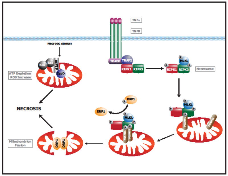
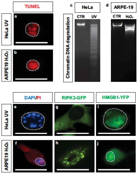
Age-related macular degeneration (AMD) is the leading cause of irreversible blindness in the elderly. The underlying mechanism of non-neovascular AMD (dry AMD), also named geographic atrophy (GA) remains unclear and the mechanism of retinal pigment epithelial (RPE) cell death in AMD is controversial. We review the history and recent progress in understanding the mechanism of RPE cell death induced by oxidative stress, in AMD mouse models, and in AMD patients. Due to the limitation of toolsets to distinguish between apoptosis and necroptosis (or necrosis), most previous research concludes that apoptosis is a major mechanism for RPE cell death in response to oxidative stress and in AMD. Recent studies suggest necroptosis as a major mechanism of RPE cell death in response to oxidative stress. Moreover, ultrastructural and histopathological studies support necrosis as major mechanism of RPE cells death in AMD. In this review, we discuss the mechanism of RPE cell death in response to oxidative stress, in AMD mouse models, and in human AMD patients. Based on the literature, we hypothesize that necroptosis is a major mechanism for RPE cell death in response to oxidative stress and in AMD.

Keywords: Age-related macular degeneration; Apoptosis; Geographic atrophy; Necroptosis; Necrosis; Oxidative stress; RPE.


Conflict of interest statement

The authors have no proprietary interest.

Figures

Figure 1
Figure 1. Overview of the apoptotic intrinsic and extrinsic pathways
Extrinsic apoptotic pathway is induced by activation of the membrane death receptors that leads to activation of the effector caspases and triggers caspase cascade that leads to cleavage of the caspases final targets, among the others: nuclear lamins, DFF45, PARP. Intrinsic pathway is triggered by DNA damage, gamma radiation, UV radiation, excessive ROS levels, virus infection or oncogenes activation. Activation of Bcl2 proteins leads to release of cytochrome c from mitochondrial intermembrane space leads to formation of apoptosome that induces activation of caspase-9 and effector caspase.
Figure 2
Figure 2. Overview of the necrotic pathways
Activation of TNF receptor leads to activation of RIP1 and RIP3 kinases when caspase-8 is not present or inactive. Autophosporylations of RIP1 and RIP3 leads to formation on necrosome, a complex that initiates necrotic signaling pathway. Necrosome recruits MLKL protein and assembles signaling complex at the membrane rafts. Recruitment of the PGAM5 marks translocation of the complex to hydrophilic environment and attachment to the mitochondrial membrane and activation of Drp1 protein that leads to mitochondrial fission and cell death. In the necrosis triggered by calcium overload, opening of mPTP leads to decrease of mitochondrial membrane potential, collapse of ATP production and release of ROS and triggering of necrosis in RIP3 dependent manner, although the signaling pathways leading to that event are unknown.
Figure 3
Figure 3. Comparison of apoptotic and necrotic hallmarks
To compare features of apoptosis and necrosis we exposed HeLa cells to UV irradiation (80J/cm2) and ARPE-19 cells to hydrogen peroxide (300μM). Analysis of DNA degradation revealed that both apoptotic HeLa cells (a) and necrotic ARPE-19 cells (b) are TUNEL positive 24 hours after induction of cell death. Although both cell types have different nuclear morphology. Additionally analysis of chromatin degradation by electrophoresis reveals organized pattern of DNA degradation known as apoptotic ladder in HeLa cells (c), while in ARPE-19 unorganized DNA degradation is visible as a smear (d). DAPI staining shows strictly orchestrated process of nuclear fragmentation in apoptotic HeLa cells (e) which is revealed in nuclear morpholoty. Additionally cell membrane of the apoptotic cells remains intact throughout the whole process therefore it is resistant to propidium iodate. Necrotic cells lose membrane integrity early and it can be crossed by PI (f) and nucleus often shrinks and disintegrates. RIP3 kinase is a switch between apoptosis and necrosis. In apoptotic cells RIP3 kinase is degraded by caspase-3 (g, picture taken one hour after exposing HeLa cells to UV irradiation, points of RIP3 aggregation are not apoptosis specific and due to RIP3 overexpression), in necrosis RIP3 kinase forms distinct punctuations reflecting its activation and formation of the necrosome (h, picture taken 1 hour after exposing ARPE-19 cells to 300μM H2O2). HMGB1 is a chromatin structural protein, in apoptosis it binds tightly to DNA and is packed together with DNA into apoptotic bodies reflecting chromatin fragmentation pattern (i). During necrosis HMGB1 is passively released from the nucleus to the cytoplasm, due to DNA modifications, through compromised nuclear envelope (j). RIP3 kinase was overexpressed as fusion protein RIPK3-GFP. HMGB1 protein was overexpressed as HMGB1-YFP fusion protein. Scale bar represents 25μm. Dashed line marks the region corresponding to the nucleus.
Figure 4
Figure 4. Model of AMD pathogenesis
RPE cells are exposed to highly oxidative environment shuffling waste from photoreceptors, and providing trophic support. As the eye ages cholesterol starts to accumulate on Bruch’s membrane, and lipofucsin packets accumulate in RPE cells. Growing drusen attract macrophages and recruit choroidal dendritic cells while drusen accumulate products of lipid peroxidation they start to increase oxidative pressure on RPE cells (A). Damaged RPE cells die from necrosis, contributing to drusen growth, promoting inflammatory response by releasing DAMP molecules and promoting damage of the neighboring RPE cells (B). Those events attribute to self-perpetuating RPE cell death events (C).

References

    1. Age-Related Eye Disease Study 2 Research G. Lutein + zeaxanthin and omega-3 fatty acids for age-related macular degeneration: the Age-Related Eye Disease Study 2 (AREDS2) randomized clinical trial. JAMA. 2013;309:2005–2015. - PubMed
    1. Age-Related Eye Disease Study Research G. A randomized, placebo-controlled, clinical trial of high-dose supplementation with vitamins C and E, beta carotene, and zinc for age-related macular degeneration and vision loss: AREDS report no. 8. Archives of ophthalmology. 2001;119:1417–1436. - PMC - PubMed
    1. Alge CS, Priglinger SG, Neubauer AS, Kampik A, Zillig M, Bloemendal H, Welge-Lussen U. Retinal pigment epithelium is protected against apoptosis by alphaB-crystallin. Invest Ophthalmol Vis Sci. 2002;43:3575–3582. - PubMed
    1. Ambati J, Anand A, Fernandez S, Sakurai E, Lynn BC, Kuziel WA, Rollins BJ, Ambati BK. An animal model of age-related macular degeneration in senescent Ccl-2- or Ccr-2-deficient mice. Nat Med. 2003;9:1390–1397. - PubMed
    1. Baich A, Ziegler M. The effect of sodium iodate and melanin on the formation of glyoxylate. Pigment cell research/sponsored by the European Society for Pigment Cell Research and the International Pigment Cell Society. 1992;5:394–395. - PubMed