

Assessment of Antiviral Properties of Peramivir against H7N9 Avian Influenza Virus in an Experimental Mouse Model
- PMID: 26369969
- PMCID: PMC4649212
- DOI: 10.1128/AAC.01885-15
Assessment of Antiviral Properties of Peramivir against H7N9 Avian Influenza Virus in an Experimental Mouse Model
Abstract
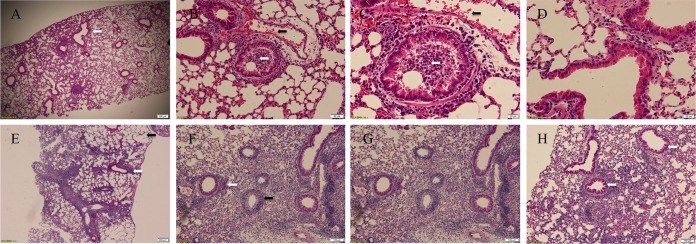
The H7N9 influenza virus causes a severe form of disease in humans. Neuraminidase inhibitors, including oral oseltamivir and injectable peramivir, are the first choices of antiviral treatment for such cases; however, the clinical efficacy of these drugs is questionable. Animal experimental models are essential for understanding the viral replication kinetics under the selective pressure of antiviral agents. This study demonstrates the antiviral activity of peramivir in a mouse model of H7N9 avian influenza virus infection. The data show that repeated administration of peramivir at 30 mg/kg of body weight successfully eradicated the virus from the respiratory tract and extrapulmonary tissues during the acute response, prevented clinical signs of the disease, including neuropathy, and eventually protected mice against lethal H7N9 influenza virus infection. Early treatment with peramivir was found to be associated with better disease outcomes.
Copyright © 2015 Farooqui et al.
Figures


Similar articles
-
The evolution and characterization of influenza A(H7N9) virus under the selective pressure of peramivir.Virology. 2019 Oct;536:58-67. doi: 10.1016/j.virol.2019.08.004. Epub 2019 Aug 6. Virology. 2019. PMID: 31400550
-
Emergence of H7N9 Influenza A Virus Resistant to Neuraminidase Inhibitors in Nonhuman Primates.Antimicrob Agents Chemother. 2015 Aug;59(8):4962-73. doi: 10.1128/AAC.00793-15. Epub 2015 Jun 8. Antimicrob Agents Chemother. 2015. PMID: 26055368 Free PMC article.
-
Inhibition of novel reassortant avian influenza H7N9 virus infection in vitro with three antiviral drugs, oseltamivir, peramivir and favipiravir.Antivir Chem Chemother. 2014 Dec 16;23(6):237-40. doi: 10.3851/IMP2672. Antivir Chem Chemother. 2014. PMID: 23948557
-
Recent advances in neuraminidase inhibitor development as anti-influenza drugs.ChemMedChem. 2012 Sep;7(9):1527-36. doi: 10.1002/cmdc.201200155. Epub 2012 Jul 16. ChemMedChem. 2012. PMID: 22807317 Review.
-
Neuraminidase inhibitor resistance in influenza viruses.J Med Virol. 2007 Oct;79(10):1577-86. doi: 10.1002/jmv.20951. J Med Virol. 2007. PMID: 17705169 Review.
Cited by
-
Peramivir injection in the treatment of acute influenza: a review of the literature.Infect Drug Resist. 2016 Aug 22;9:201-14. doi: 10.2147/IDR.S86460. eCollection 2016. Infect Drug Resist. 2016. PMID: 27578993 Free PMC article. Review.
-
Peramivir: A Novel Intravenous Neuraminidase Inhibitor for Treatment of Acute Influenza Infections.Front Microbiol. 2016 Mar 31;7:450. doi: 10.3389/fmicb.2016.00450. eCollection 2016. Front Microbiol. 2016. PMID: 27065996 Free PMC article. Review.
-
Inhibition of avian-origin influenza A(H7N9) virus by the novel cap-dependent endonuclease inhibitor baloxavir marboxil.Sci Rep. 2019 Mar 5;9(1):3466. doi: 10.1038/s41598-019-39683-4. Sci Rep. 2019. PMID: 30837531 Free PMC article.
-
Pathogenicity and peramivir efficacy in immunocompromised murine models of influenza B virus infection.Sci Rep. 2017 Aug 4;7(1):7345. doi: 10.1038/s41598-017-07433-z. Sci Rep. 2017. PMID: 28779075 Free PMC article.
References
-
- Chen E, Chen Y, Fu L, Chen Z, Gong Z, Mao H, Wang D, Ni M, Wu P, Yu Z, He T, Li Z, Gao J, Liu S, Shu Y, Cowling BJ, Xia S, Yu H. 2013. Human infection with avian influenza A (H7N9) virus re-emerges in China in winter 2013. Euro Surveill 18:20616. - PubMed
-
- WHO. 2015. Human infection with avian influenza A(H7N9) virus—China. World Health Organization, Geneva, Switzerland.
-
- China Health and Family Planning Commission. 2015. National statutory infectious diseases overview. China Health and Family Planning Commission, Beijing, China.
Publication types
MeSH terms
Substances
LinkOut - more resources
Full Text Sources
Medical
