. 2016 Feb;30(2):492-500.
doi: 10.1038/leu.2015.247. Epub 2015 Sep 15.

Chimeric antigen receptor-modified T cells derived from defined CD8+ and CD4+ subsets confer superior antitumor reactivity in vivo

Affiliations

Chimeric antigen receptor-modified T cells derived from defined CD8+ and CD4+ subsets confer superior antitumor reactivity in vivo

D Sommermeyer et al. Leukemia. 2016 Feb.

Abstract

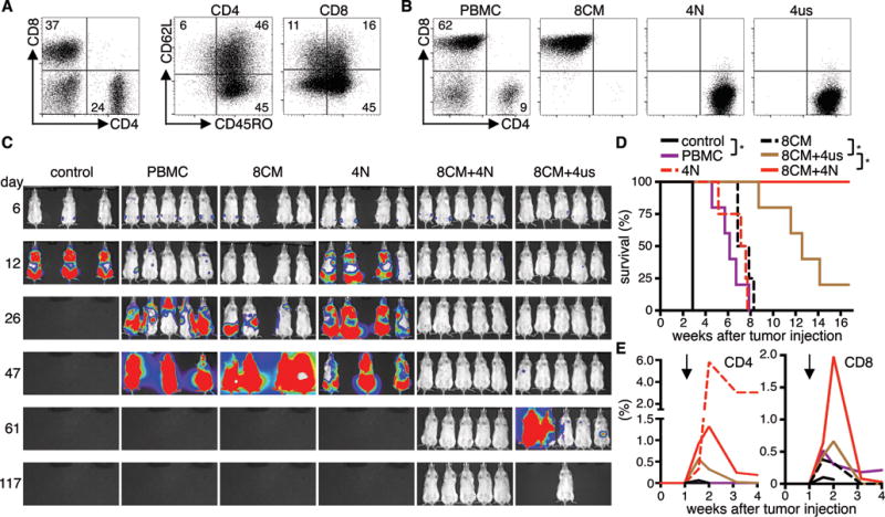
Adoptive T-cell therapy with gene-modified T cells expressing a tumor-reactive T-cell receptor or chimeric antigen receptor (CAR) is a rapidly growing field of translational medicine and has shown success in the treatment of B-cell malignancies and solid tumors. In all reported trials, patients have received T-cell products comprising random compositions of CD4(+) and CD8(+) naive and memory T cells, meaning that each patient received a different therapeutic agent. This variation may have influenced the efficacy of T-cell therapy, and complicates comparison of outcomes between different patients and across trials. We analyzed CD19 CAR-expressing effector T cells derived from different subsets (CD4(+)/CD8(+) naive, central memory, effector memory). T cells derived from each of the subsets were efficiently transduced and expanded, but showed clear differences in effector function and proliferation in vitro and in vivo. Combining the most potent CD4(+) and CD8(+) CAR-expressing subsets, resulted in synergistic antitumor effects in vivo. We show that CAR-T-cell products generated from defined T-cell subsets can provide uniform potency compared with products derived from unselected T cells that vary in phenotypic composition. These findings have important implications for the formulation of T-cell products for adoptive therapies.


Conflict of interest statement

Conflict of interest statement:

M.H. and S.R.R. are inventors on a patent application (PCT/US1013/055862) related to this work that has been filed by the Fred Hutchinson Cancer Research Center (FHCRC) and licensed by Juno Therapeutics. S.R.R. is founder and shareholder of Juno Therapeutics.

Figures

Figure 1
Figure 1. T-cell subset composition in PBMC from patients and healthy donors
Proportions of (A) CD4+ and CD8+ T-cells, and (B) TN, TCM, and TEM cell subsets in blood of patients with B-cell malignancies and normal donors. Asterisk indicates significant differences between normal donors and patients.
Figure 2
Figure 2. CD4+ and CD8+ CAR-T-cells differ in effector function
(A) Expression of EGFRt on CD4+ and CD8+ T-cells transduced with a CD19 CAR-EGFRt construct before enrichment (pre) and after enrichment and expansion (post). (B) Cytolytic activity of CD19 CAR-T-cells against 51Cr-labeled CD19+ (K562/CD19, Raji) and control (K562) target cells at an E:T ratio of 30:1 analyzed by a standard 4 h chromium release assay. (C) Proliferation of CFSE-labeled CD19 CAR-T-cells after stimulation with K562/CD19 and Raji cells for 72 h measured by CFSE dye dilution. Stimulation of CFSE-labeled CD19 CAR-T-cells with CD19 K562 cells is shown as a comparison in each histogram (gray graph). Numbers above each histogram indicate the number of cell divisions the proliferating subset underwent. Data in A–C are representative of experiments with CAR-T-cells derived from three different donors. (D) Relative cytokine production by CD19 CAR-T-cells after co-culture with K562/CD19 and Raji cells for 24 h. Data from three independent experiments with T-cells prepared from different donors were combined. Cytokine production of CD4+ cells was set as 1 and relative cytokine production of CD8+ T-cells was calculated. Asterisk indicates significant differences between CD4+ and CD8+ cells.
Figure 3
Figure 3. In vitro and in vivo analysis of CD19 CAR-expressing naïve and memory CD4+ T-cell subsets
(A) CD4+ TN, TCM, and TEM cells were isolated by FACS. (B) Analysis of cell surface expression of CD45RO, CD62L, CD27, and CD28 on CD4+ CD19 CAR-T-cells derived from different subsets. Data from three independent experiments with T-cells prepared from different donors were combined. (C) Cytolytic activity of CAR-T-cells against 51Cr-labeled K562, K562/CD19, and Raji cells at an E:T ratio of 30:1 after 4 h. (D) Relative cytokine production by CD19 CAR-T-cells after co-culture with CD19+ cells for 24 h. Data from three independent experiments with T-cells prepared from different donors were combined. Cytokine production of TN was set as 1 and relative cytokine production of TCM and TEM was calculated. (E) Survival of Raji/ffluc-bearing NSG mice treated on day seven after tumor injection with CD19 CAR-T-cells derived from different CD4+ T-cell subsets (2.5×10⁶ or 5×10⁶ CAR-T-cells per mouse). NSG mice that received 5×10⁶ T-cells transduced with a vector encoding EGFRt alone (EGFRt-T-cells) were used as control. Asterisk indicates significant differences between groups (n.s.: not significant). Data from three independent experiments for the lower cell dose (2.5×10⁶) and two for the higher cell dose (5×10⁶) with minimum three mice per group were combined. (F) Frequency of transferred T-cells (human CD45+/CD4+) on day ten after T-cell transfer in peripheral blood of mice that had received the lower T-cell dose.
Figure 4
Figure 4. In vitro and in vivo analysis of CD19 CAR-expressing naïve and memory CD8+ T-cell subsets
(A) Analysis of cell surface expression of CD45RO, CD62L, CD27, and CD28 on CD8+ CD19 CAR-T-cells derived from naïve and memory subsets. Data from three independent experiments with T-cells prepared from different donors were combined. (B) Cytolytic activity of CD19 CAR-T-cells against 51Cr-labeled K562, K562/CD19, and Raji cells at an E:T ratio of 30:1 after 4 h. (C) Relative cytokine production by CD19 CAR-T-cells after co-culture with CD19+ cells for 24 h. Data from three independent experiments with T-cells prepared from different donors were combined. Cytokine production of TN was set as 1 and relative cytokine production of TCM and TEM was calculated. (D) Survival of Raji/ffluc-bearing NSG mice treated on day seven after tumor injection with different doses of CD19 CAR-T-cells derived from different CD8+ T-cell subsets (1×10⁶ and 2.5×10⁶). NSG mice that received 2.5×10⁶ EGFRt-T-cells were used as control. Asterisk indicates significant differences between groups (n.s.: not significant). For the lower cell dose (1×10⁶) four and for the higher cell dose (2.5×10⁶) three experiments with minimum three mice per group are combined. (E) Frequency of transferred T-cells (human CD45+/CD8+) on day ten after T-cell transfer in peripheral blood of mice that had received the lower T-cell dose.
Figure 5
Figure 5. CD4+ CD19 CAR-T-cells provide help to CD8+ CD19 CAR-T-cells in vitro and in vivo
(A) Proliferation of CFSE-labeled CD8+ TCM-derived CD19 CAR-T-cells after stimulation with JeKo-1 and Raji cells for 72 h in the presence of CD4+ CD19 CAR-T-cells derived from different subsets. Histograms are gated on CD8+ T-cells. Stimulation with K562 cells is shown as a comparison in each histogram (gray graph). Data are representative for four independent experiments with T-cells generated from different healthy donors. (B) Cytokine production of CD4+ CD19 CAR-T-cells derived from different subsets from a CLL-patient after co-culture with K562/CD19 for 24 h. (C) Same as (A) with T-cells from a CLL-patient and K562/CD19 cells and autologous CLL as stimulator cells in the assay. Data in (B) and (C) are representative for two independent experiments with T-cells generated from different patients. (D) Survival of Raji/ffluc-bearing NSG mice treated on day seven after tumor injection with 8×10⁵ CD19 CAR-T-cells derived from CD4+ TN, TCM, TEM, and CD8+ TCM (CD4/CD8 combinations: 4×10⁵ CD4+ cells + 4×10⁵ CD8+ cells; 5 mice per group). NSG mice that received a combination of CD4+ and CD8+ EGFRt-T-cells were used as control. (E) Bioluminescence imaging of tumor growth. Arrows mark the day of T-cell transfer. (F) Frequency of transferred CD4+ and CD8+ T-cells (human CD45+/CD4+ or CD45+/CD8+) in peripheral blood. (G) Flow cytometry data on day ten after T-cell transfer. Mice with median T-cell frequency are shown for each group.
Figure 6
Figure 6. T-cell products of defined subset composition prepared from patients have enhanced potency
(A) T-cell subset composition in PBMC from a lymphoma patient. (B) CD4/CD8 phenotype of CD19 CAR-T-cell products derived from PBMC or from different sort-purified subsets. (C) Bioluminescence imaging data from Raji/ffluc-bearing NSG mice that had received 1×10⁶ CD19 CAR-T-cells (CD4/CD8 ratio: 1:1, 4–5 mice per group). NSG mice that received EGFRt-T-cells were used as control (3 mice). (D) Survival and (E) persistence of transferred CD4+ and CD8+ T-cells in the blood of mice shown in (C). Asterisk indicates significant differences between groups. Arrows mark the day of T-cell transfer. Data are representative for two independent experiments with T-cells generated from different patients.

References

    1. Jensen MC, Riddell SR. Design and implementation of adoptive therapy with chimeric antigen receptor-modified T cells. Immunol Rev. 2014 Jan;257(1):127–144. - PMC - PubMed
    1. Kochenderfer JN, Rosenberg SA. Treating B-cell cancer with T cells expressing anti-CD19 chimeric antigen receptors. Nature reviews Clinical oncology. 2013 May;10(5):267–276. - PMC - PubMed
    1. Davila ML, Riviere I, Wang X, Bartido S, Park J, Curran K, et al. Efficacy and Toxicity Management of 19-28z CAR T Cell Therapy in B Cell Acute Lymphoblastic Leukemia. Sci Transl Med. 2014 Feb 19;6(224):224ra225. - PMC - PubMed
    1. Grupp SA, Kalos M, Barrett D, Aplenc R, Porter DL, Rheingold SR, et al. Chimeric antigen receptor-modified T cells for acute lymphoid leukemia. N Engl J Med. 2013 Apr 18;368(16):1509–1518. - PMC - PubMed
    1. Kalos M, Levine BL, Porter DL, Katz S, Grupp SA, Bagg A, et al. T cells with chimeric antigen receptors have potent antitumor effects and can establish memory in patients with advanced leukemia. Sci Transl Med. 2011 Aug 10;3(95):95ra73. - PMC - PubMed

Publication types

Substances