. 2015 Sep 14;4(9):e002232.
doi: 10.1161/JAHA.115.002232.

Persistently Altered Brain Mitochondrial Bioenergetics After Apparently Successful Resuscitation From Cardiac Arrest

Affiliations

Persistently Altered Brain Mitochondrial Bioenergetics After Apparently Successful Resuscitation From Cardiac Arrest

Todd J Kilbaugh et al. J Am Heart Assoc. .

Abstract

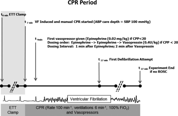
Background: Although advances in cardiopulmonary resuscitation have improved survival from cardiac arrest (CA), neurologic injury persists and impaired mitochondrial bioenergetics may be critical for targeted neuroresuscitation. The authors sought to determine if excellent cardiopulmonary resuscitation and postresuscitation care and good traditional survival rates result in persistently disordered cerebral mitochondrial bioenergetics in a porcine pediatric model of asphyxia-associated ventricular fibrillation CA.

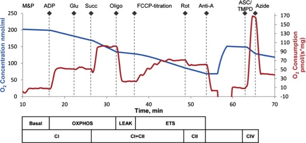
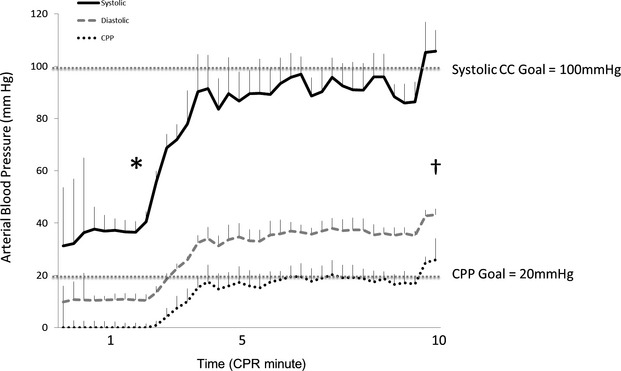
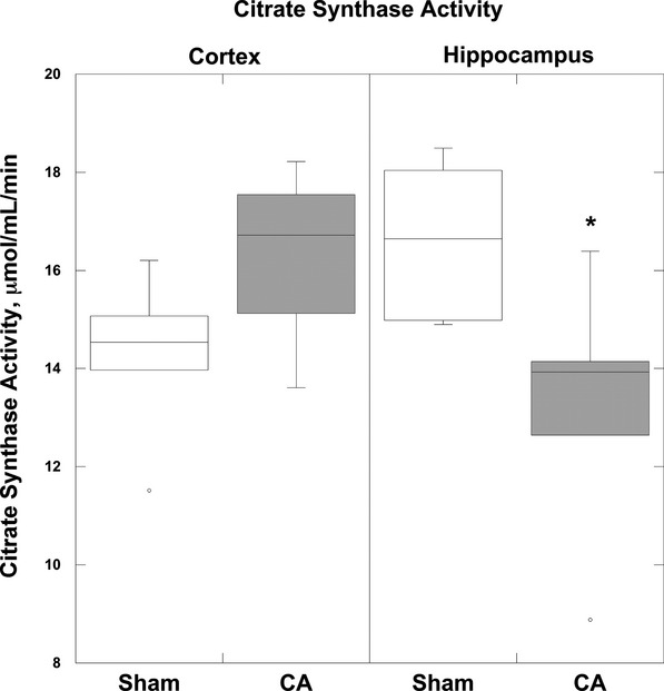
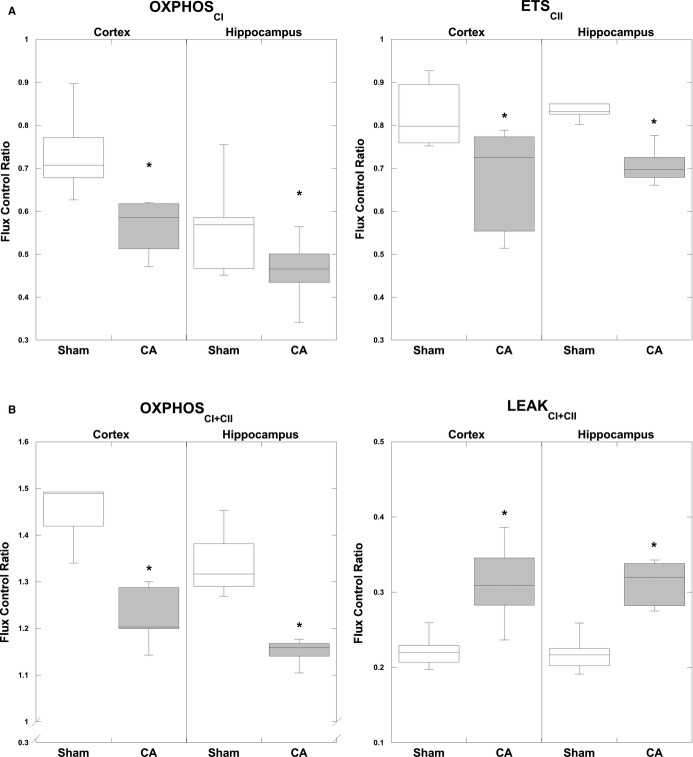
Methods and results: After 7 minutes of asphyxia, followed by ventricular fibrillation, 5 female 1-month-old swine (4 sham) received blood pressure-targeted care: titration of compression depth to systolic blood pressure of 90 mm Hg and vasopressor administration to a coronary perfusion pressure >20 mm Hg. All animals received protocol-based vasopressor support after return of spontaneous circulation for 4 hours before they were killed. The primary outcome was integrated mitochondrial electron transport system (ETS) function. CA animals displayed significantly decreased maximal, coupled oxidative phosphorylating respiration (OXPHOSCI + CII) in cortex (P<0.02) and hippocampus (P<0.02), as well as decreased phosphorylation and coupling efficiency (cortex, P<0.05; hippocampus, P<0.05). Complex I- and complex II-driven respiration were both significantly decreased after CA (cortex: OXPHOSCI P<0.01, ETSCII P<0.05; hippocampus: OXPHOSCI P<0.03, ETSCII P<0.01). In the hippocampus, there was a significant decrease in maximal uncoupled, nonphosphorylating respiration (ETSCI + CII), as well as a 30% reduction in citrate synthase activity (P<0.04).

Conclusions: Mitochondria in both the cortex and hippocampus displayed significant alterations in respiratory function after CA despite excellent cardiopulmonary resuscitation and postresuscitation care in asphyxia-associated ventricular fibrillation CA. Analysis of integrated ETS function identifies mitochondrial bioenergetic failure as a target for goal-directed neuroresuscitation after CA. IACUC Protocol: IAC 13-001023.

Keywords: acute brain injury; brain; cardiac arrest; electron transport system; mitochondria; neuroprotection.


Figures

Figure 1
Figure 1
Protocol design. Shaded area denotes period of “acute respiratory insufficiency,” induced by endotracheal tube clamping. During the CPR period, animals received chest compressions with depth titrated to an SBP of 90 mm Hg and vasopressors when the CPP was <20 mm Hg. ABP indicates arterial blood pressure; CPP, coronary perfusion pressure; CPR, cardiopulmonary resuscitation; ETT, endotracheal tube; ROSC, return of spontaneous circulation; SBP, systolic blood pressure; VF, ventricular fibrillation.
Figure 2
Figure 2
Oxygen concentration and consumption example trace from the SUIT protocol. Mitochondrial respiratory states and respiratory complexes are outlined below the x-axis. The chamber was opened to increase oxygen concentration to 150 nmol/mL before the addition of TMPD. Anti-A indicates antimycin-A; ASC/TMPD, ascorbate and N,N,N′,N′-tetramethyl-p-phenylenediamine; ETS, electron transport system; FCCP, carbonyl cyanide p-(trifluoromethoxy)phenylhydrazone; Glu, glutamate; M&P, malate+pyruvate; Oligo, oligomycin; OXPHOS, oxidative phosphorylation; Rot, rotenone; Succ, succinate; SUIT, substrate, uncoupler, inhibitor titration.
Figure 3
Figure 3
Hemodynamic resuscitation showing systolic, diastolic, and coronary perfusion pressures during CPR period. *First vasopressor dose. First shock. Error bars represent SEM. CC indicates chest compression; CPP, coronary perfusion pressure; CPR, cardiopulmonary resuscitation.
Figure 4
Figure 4
Citrate synthase activity. Four hours after CA there was a significant decrease in CS activity in the hippocampus compared with sham animals (*P<0.01). The cortex did not exhibit a significant reduction in CS activity post CA. Boxplots: Horizontal lines represent the median CS activity, with the boxes representing the 25th and 75th percentiles, the whiskers representing the 5th and 95th percentiles, and the dots representing the outlierss. CA indicates cardiac arrest; CS, citrate synthase.
Figure 5
Figure 5
Mitochondrial FCR normalized by ETS capacity (ETSCI+CII) measured 4 hours post CA. A, CA resulted in a significant decrease in complex I–driven respiration (OXPHOSCI) in both regions: cortex and hippocampus. Complex II–driven respiration (ETSCII) in CA tissue is significantly decreased bilaterally, compared with corresponding sham tissue. B, Maximal coupled, phosphorylating respiration (OXPHOSCI+CII), stimulated by both complex I and complex II substrates, was significantly decreased post CA in both regions compared with shams. State 4o (LEAKCI+CII) displayed a significant increase 4 hours post CA compared with shams in the cortex and the hippocampus. For definitions of respiratory states and substrates, please see Figure 1. *P<0.05. Boxplots: Horizontal lines represent the median FCR, with the boxes representing the 25th and 75th percentiles, the whiskers representing the 5th and 95th percentiles, and the dots representing the outliers. CA indicates cardiac arrest; ETS, electron transport system; FCR, flux control ratios; OXPHOSCI+CII, oxidative phosphorylation capacity of complexes I and II.

References

    1. Merchant RM, Yang L, Becker LB, Berg RA, Nadkarni V, Nichol G, Carr BG, Mitra N, Bradley SM, Abella BS, Groeneveld PW American Heart Association Get With The Guidelines-Resuscitation Investigators. Incidence of treated cardiac arrest in hospitalized patients in the United States. Crit Care Med. 2011;39:2401–2406. - PMC - PubMed
    1. Girotra S, Chan PS. Trends in survival after in-hospital cardiac arrest. N Engl J Med. 2013;368:680–681. - PubMed
    1. Ayoub IM, Radhakrishnan J, Gazmuri RJ. Targeting mitochondria for resuscitation from cardiac arrest. Crit Care Med. 2008;36:S440–S446. - PMC - PubMed
    1. Gazmuri RJ, Radhakrishnan J. Protecting mitochondrial bioenergetic function during resuscitation from cardiac arrest. Crit Care Clin. 2012;28:245–270. - PMC - PubMed
    1. Han F, Da T, Riobo NA, Becker LB. Early mitochondrial dysfunction in electron transfer activity and reactive oxygen species generation after cardiac arrest. Crit Care Med. 2008;36:S447–S453. - PMC - PubMed

Publication types

MeSH terms

Substances