. 2015 Dec;35(23):3962-73.
doi: 10.1128/MCB.00813-15. Epub 2015 Sep 14.

High CO2 Leads to Na,K-ATPase Endocytosis via c-Jun Amino-Terminal Kinase-Induced LMO7b Phosphorylation

Affiliations

High CO2 Leads to Na,K-ATPase Endocytosis via c-Jun Amino-Terminal Kinase-Induced LMO7b Phosphorylation

Laura A Dada et al. Mol Cell Biol. 2015 Dec.

Abstract

The c-Jun amino-terminal kinase (JNK) plays a role in inflammation, proliferation, apoptosis, and cell adhesion and cell migration by phosphorylating paxillin and β-catenin. JNK phosphorylation downstream of AMP-activated protein kinase (AMPK) activation is required for high CO2 (hypercapnia)-induced Na,K-ATPase endocytosis in alveolar epithelial cells. Here, we provide evidence that during hypercapnia, JNK promotes the phosphorylation of LMO7b, a scaffolding protein, in vitro and in intact cells. LMO7b phosphorylation was blocked by exposing the cells to the JNK inhibitor SP600125 and by infecting cells with dominant-negative JNK or AMPK adenovirus. The knockdown of the endogenous LMO7b or overexpression of mutated LMO7b with alanine substitutions of five potential JNK phosphorylation sites (LMO7b-5SA) or only Ser-1295 rescued both LMO7b phosphorylation and the hypercapnia-induced Na,K-ATPase endocytosis. Moreover, high CO2 promoted the colocalization and interaction of LMO7b and the Na,K-ATPase α1 subunit at the plasma membrane, which were prevented by SP600125 or by transfecting cells with LMO7b-5SA. Collectively, our data suggest that hypercapnia leads to JNK-induced LMO7b phosphorylation at Ser-1295, which facilitates the interaction of LMO7b with Na,K-ATPase at the plasma membrane promoting the endocytosis of Na,K-ATPase in alveolar epithelial cells.


Figures

FIG 1
FIG 1
LMO7b is phosphorylated during hypercapnia in T/S-P domain. (A) rATII cells were exposed to 120 mm Hg CO2 for 10 min in the presence or absence of the inhibitor of JNK phosphorylation, SP600125, and analyzed by Western blotting (WB) using a pT/S-P antibody. A band at ∼150 kDa was detected only in the absence of SP600125. The nLC MS/MS analysis of the band at ∼150 kDa excised from a parallel gel identified peptides corresponding to rat LMO7b. Calc., calculated; Obsrv., observed. (B) Expression of LMO7b isoforms in isolated rATII cells. (C) rATII cells were exposed for 10 min to either normocapnia (40 mm Hg CO2) or increasing concentrations of CO2. LMO7b was immunoprecipitated, and phosphorylation was assessed using a pT/S-P by WB. Immunoprecipitated LMO7b was used as a loading control. Results are means ± SEM; n = 3. (D) rATII cells were exposed to hypercapnia (120 mm Hg CO2) for the indicated times. Samples were processed as described for panel C. Results are means ± SEM; n = 3. (E) A549 cells expressing rat FLAG-LMO7b were exposed to 40 mm Hg CO2 (CT) or 120 mm Hg CO2 (CO2) for 10 min. LMO7b was immunoprecipitated using a FLAG antibody. Phosphorylation was assessed by WB using a pT/S-P or a pMAPK substrate (pMAPK) antibody. FLAG was used as a loading control. *, P < 0.05; **, P < 0.01.
FIG 2
FIG 2
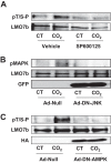
LMO7b is phosphorylated during hypercapnia downstream of AMPK and JNK. (A) rATII cells were pretreated with SP600125 (20 μM for 30 min) and then exposed to normocapnia (CT) or hypercapnia (CO2) for 10 min. LMO7b was immunoprecipitated and phosphorylation assessed by WB with a pT/S-P antibody. Immunoprecipitated LMO7b is shown as a loading control. (B and C) rATII cells were infected with a null adenovirus (Ad-null) or an adenovirus expressing DN-JNK and GFP (B) or with Ad-null or Ad-DN-HA-AMPK (C). After 24 h, cells were exposed to CO2 and processed as described for panel A, and WB were developed with a pMAPK (B) or with a pT/S-P antibody (C). GFP and HA Western blots are shown as markers of infection. n = 3.
FIG 3
FIG 3
LMO7b is a target for JNK phosphorylation. (A) In vitro JNK kinase assay was performed by incubating purified recombinant JNK1 (150 ng) with LMO7b immunoprecipitated (IP) from rATII cells in the presence or absence of [γ-32P]ATP. A representative autoradiograph of phosphorylated LMO7b (32P) and Western blot (WB) of LMO7b are shown. n = 3. (B) In vitro JNK kinase assay using rat FLAG-LMO7b as a substrate immunoprecipitated from A549 transfected cells. n = 3. (C) In vitro kinase assay was performed in the presence or absence of SP600125 (20 μM) as described for panel A. A representative autoradiograph of phosphorylated LMO7b (32P) and WB analysis of LMO7b are shown. n = 3. (D) A schematic depicting rat LMO7b potential JNK phosphorylation sites. (E) In vitro JNK kinase assay was performed as described for panel A using LMO7b-FLAG (WT) or FLAG-LMO7b bearing a serine-to-alanine mutation in positions 727, 1295, 1298, 1300, and 1305 (FLAG-LMO7b-5SA). A representative autoradiograph of phosphorylated LMO7 (32P) and WB analysis of LMO7 using FLAG antibody are shown. n = 3. (F) A549 cells were transfected with FLAG-LMO7b-WT or FLAG-LMO7b-5SA, and 48 h later cells were exposed for 10 min to either normocapnia (CT) or hypercapnia (CO2). LMO7b was immunoprecipitated using FLAG antibody, and the phosphorylation of LMO7 was analyzed by WB using a pT/S-P antibody. The total amount of immunoprecipitated LMO7b was measured using FLAG antibody. n = 3.
FIG 4
FIG 4
Hypercapnia-induced Na,K-ATPase endocytosis requires LMO7b phosphorylation. (A) A549 cells were transfected with an siRNA against LMO7b (si LMO7b) or scrambled siRNA (si scr) and exposed for 30 min to either normocapnia (CT) or hypercapnia (CO2). Na,K-ATPase at the plasma membrane was determined by biotin-streptavidin pulldown and Western blotting (WB). Representative WB of Na,K-ATPase-α1 and LMO7 are shown. Values are expressed as means ± SEM. n = 3. (B) Schematic depicting the different FLAG-LMO7b constructs used. (C) A549 cells were transfected with FLAG-LMO7b-WT, FLAG-LMO7b-5SA, FLAG-LMO7b-4SA, and FLAG-LMO7b-3SA, and 48 h later cells were exposed for 10 min to either normocapnia or hypercapnia. Na,K-ATPase expression at the plasma membrane was performed as described for panel A. Values are expressed as means ± SEM. n = 3. *, P < 0.05; **, P < 0.01.
FIG 5
FIG 5
LMO7b is phosphorylated at Ser-1295 during hypercapnia. (A) A549 cells were transfected at FLAG-LMO7b-WT, FLAG-LMO7b-S727A-S1305A, FLAG-LMO7b-S727A-S1295A, FLAG-LMO7b-S727A, or FLAG-LMO7b-S1295A, and 48 h later cells were exposed for 30 min to either normocapnia (CT) or hypercapnia (CO2). Na,K-ATPase expression at the plasma membrane was determined by biotin-streptavidin pulldown and Western blotting (WB). Representative WB of Na,K-ATPase-α1 and FLAG are shown. Values are expressed as means ± SEM. n = 3. (B) In vitro JNK kinase assay was performed by incubating purified recombinant JNK1 (150 ng) with FLAG-LMO7b-WT, FLAG-LMO7b-S727A, FLAG-LMO7b-S1295A, FLAG-LMO7b-S1300A, and FLAG-LMO7b-S1305A immunoprecipitated from A549 cells in the presence or absence of [γ-32P]ATP. A representative autoradiography of phosphorylated LMO7b (32P) and WB of FLAG-LMO7b are shown. (Right) Quantification of the phosphorylation levels in the different immunoprecipitated mutants. Values shown are means ± SEM. n = 3. *, P < 0.05; **, P < 0.01.
FIG 6
FIG 6
LMO7b colocalizes with Na,K-ATPase during hypercapnia. (A) Immunofluorescence images of rATII cells exposed for 15 min to either normocapnia (CT) or hypercapnia (CO2). The merged images were assessed by indirect multichannel acquisition. The green channel represents Na,K-ATPase-α1, the red channel represents LMO7b, and yellow indicates colocalization. z-stacks were collected at stepwise z increments from the basal to the apical side of the cell. (B) Immunofluorescence images of rATII cells exposed for 15 min to either normocapnia or hypercapnia in the presence or absence of SP600125 (SP; 20 μM for 30 min). The merged images were assessed by indirect multichannel acquisition. The green channel represents Na,K-ATPase-α1, the red channel represents LMO7b, and yellow indicates colocalization. (C) Quantification of the colocalization of Na,K-ATPase-α1 and LMO7b. At least 7 images where analyzed for each condition. *, P < 0.05; **, P < 0.01.
FIG 7
FIG 7
JNK-induced phosphorylation of LMO7b at Ser-1295 is required for Na,K-ATPase-LMO7b interaction during hypercapnia. (A) A549-GFP-α1 cells were transfected with LMO7b-FLAG-WT, and 48 h later cells were exposed for 15 min to either normocapnia (CT) or hypercapnia (CO2) in the presence of SP600125 (20 μM; 30-min preincubation) or dimethyl sulfoxide (DMSO) (vehicle). Cells were lysed using a low-detergent buffer, and Na,K-ATPase-α1 subunit was immunoprecipitated (IP) using GFP antibody. Representative WB showing Na,K-ATPase-α1 subunit or LMO7b are shown. n = 3. (B) A549-GFP-α1 cells were transfected with FLAG-LMO7b-WT or FLAG-LMO7b-S1295A, and 48 h later cells were exposed for 15 min to either normocapnia or hypercapnia and processed as described for panel A. n = 3.
FIG 8
FIG 8
Hypercapnia increases the interaction of LMO7b with endocytic proteins. (A) A549-GFP-α1 cells were transfected with LMO7b-FLAG-WT, and 48 h later cells were exposed for 15 min to either normocapnia (CT) or hypercapnia (CO2). Cells were lysed using a low-detergent buffer, and Na,K-ATPase-α1 subunit was immunoprecipitated (IP) using GFP antibody. Representative WB showing Na,K-ATPase-α1 subunit or LMO7b are shown. n = 3. (B) rATII cells were exposed to normocapnia or hypercapnia for 15 min. Cells were lysed as described for panel A, and LMO7b was immunoprecipitated with the LMO7b M300 antibody and processed as described in Materials and Methods. The table depicts proteins that coimmunoprecipitated with LMO7b as analyzed by nLC MS/MS. Endocytosis-related proteins are shown in boldface. n = 3.

Similar articles

Cited by

References

    1. Kessler R, Faller M, Fourgaut G, Mennecier B, Weitzenblum E. 1999. Predictive factors of hospitalization for acute exacerbation in a series of 64 patients with chronic obstructive pulmonary disease. Am J Respir Crit Care Med 159:158–164. doi:10.1164/ajrccm.159.1.9803117. - DOI - PubMed
    1. Vitacca M, Bianchi L, Barbano L, Ziliani M, Ambrosino N. 2005. Effects of acute on chronic respiratory failure on hypercapnia and 3-month survival. Chest 128:1209–1215. doi:10.1378/chest.128.3.1209. - DOI - PubMed
    1. Vadasz I, Hubmayr RD, Nin N, Sporn PH, Sznajder JI. 2012. Hypercapnia: a nonpermissive environment for the lung. Am J Respir Cell Mol Biol 46:417–421. doi:10.1165/rcmb.2011-0395PS. - DOI - PMC - PubMed
    1. Kohnlein T, Windisch W, Wegscheider K, Welte T. 2014. Non-invasive positive pressure ventilation for severe COPD. Authors' reply. Lancet Respir Med 2:e19. - PubMed
    1. Briva A, Vadasz I, Lecuona E, Welch LC, Chen J, Dada LA, Trejo HE, Dumasius V, Azzam ZS, Myrianthefs PM, Batlle D, Gruenbaum Y, Sznajder JI. 2007. High CO2 levels impair alveolar epithelial function independently of pH. PLoS One 2:e1238. doi:10.1371/journal.pone.0001238. - DOI - PMC - PubMed

Publication types

MeSH terms