. 2015 Sep 15;17(1):249.
doi: 10.1186/s13075-015-0771-6.

Apremilast, a novel phosphodiesterase 4 (PDE4) inhibitor, regulates inflammation through multiple cAMP downstream effectors

Affiliations

Apremilast, a novel phosphodiesterase 4 (PDE4) inhibitor, regulates inflammation through multiple cAMP downstream effectors

Miguel Perez-Aso et al. Arthritis Res Ther. .

Abstract

Introduction: This work was undertaken to delineate intracellular signaling pathways for the PDE4 inhibitor apremilast and to examine interactions between apremilast, methotrexate and adenosine A2A receptors (A2AR).

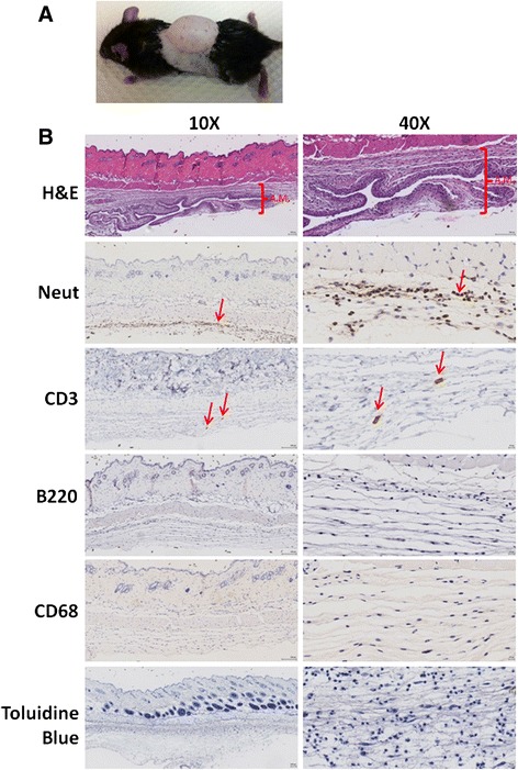
Methods: After apremilast and LPS incubation, intracellular cAMP, TNF-α, IL-10, IL-6 and IL-1α were measured in the Raw264.7 monocytic murine cell line. PKA, Epac1/2 (signaling intermediates for cAMP) and A2AR knockdowns were performed by shRNA transfection and interactions with A2AR and A2BR, as well as with methotrexate were tested in vitro and in the murine air pouch model. Statistical differences were determined using one or two-way ANOVA or Student's t test. The alpha nominal level was set at 0.05 in all cases. A P value of < 0.05 was considered significant.

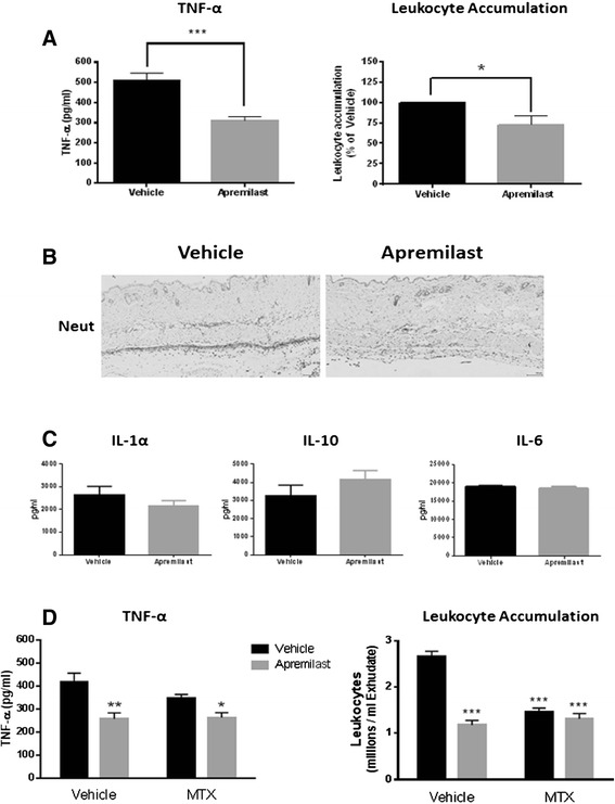
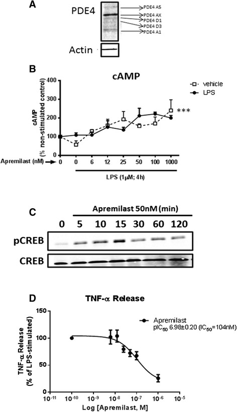
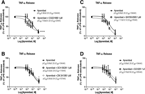
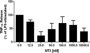
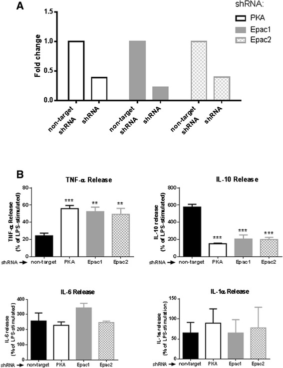
Results: In vitro, apremilast increased intracellular cAMP and inhibited TNF-α release (IC50=104nM) and the specific A2AR-agonist CGS21680 (1μM) increased apremilast potency (IC50=25nM). In this cell line, apremilast increased IL-10 production. PKA, Epac1 and Epac2 knockdowns prevented TNF-α inhibition and IL-10 stimulation by apremilast. In the murine air pouch model, both apremilast and MTX significantly inhibited leukocyte infiltration, while apremilast, but not MTX, significantly inhibited TNF-α release. The addition of MTX (1 mg/kg) to apremilast (5 mg/kg) yielded no more inhibition of leukocyte infiltration or TNF-α release than with apremilast alone.

Conclusions: The immunoregulatory effects of apremilast appear to be mediated by cAMP through the downstream effectors PKA, Epac1, and Epac2. A2AR agonism potentiated TNF-α inhibition by apremilast, consistent with the cAMP-elevating effects of that receptor. Because the A2AR is also involved in the anti-inflammatory effects of MTX, the mechanism of action of both drugs involves cAMP-dependent pathways and is therefore partially overlapping in nature.


Figures

Fig. 1
Fig. 1
Air pouch model of inflammation. a Inflammation in the air pouch was induced as described. b H&E reveals the formation of the air pouch membrane (A.M.) and immunohistology staining with specific neutrophil (Neut), T cell (CD3), B cell (B220) and macrophage (CD68) markers, and the toluidine blue stain for mast cells was performed
Fig. 2
Fig. 2
Apremilast and methotrexate (MTX) prevent inflammation in the air pouch independently. Mice were orally treated with apremilast (5 mg/Kg), and inflammation in the air pouch was induced as described under “Materials and methods”. a TNF-α and leukocyte accumulation were quantified in the exudates of the air pouch. b Immunohistology reveals a decrease in the number of neutrophils (Neut) by apremilast treatment. c IL-1α, IL-6 and IL-10 levels were measured in the air pouch exudates with the Luminex multiplex technology. d TNF-α and leukocyte were quantified in the air pouch exudates in mice after weekly intraperitoneal injections of MTX (1 mg/Kg) for 4 weeks prior to apremilast treatment. Data represent mean ± standard error of the mean of at least three independent experiments. Statistical analysis was performed by the Student’s t test or one-way ANOVA, where ***P <0.001, **P <0.01 and *P <0.05 vs vehicle
Fig. 3
Fig. 3
Apremilast inhibits lipopolysaccharide (LPS)-induced TNF-α release via cyclic adenosine monophosphalphate (cAMP). a Western blot of PDE4 was performed in the Raw 264.7 cell line. b Raw 264.7 cells were incubated with increasing concentrations of apremilast 30 minutes before incubation with or without LPS 1 μM during 20 minutes. Intracellular cAMP levels were then measured as described under “Materials and methods”. c Western blot of p-cAMP responsive element binding protein (p-CREB) and CREB shows that apremilast promotes CREB phosphorylation after incubation with LPS 1 μg/ml for 30 minutes. d Cumulative concentration response curves to apremilast (6 nM−1 μM) were performed in the Raw 264.7 cells 30 minutes before incubation with LPS 1 μM during 4 h. IC50 values were determined as described under “Materials and methods”. Data represent means ± standard error of the mean of four independent experiments. Statistical analysis was performed by two-way ANOVA: apremilast ***P <0.001, LPS: not significant. PDE4 phosphodiesterase 4,
Fig. 4
Fig. 4
Combined effects adenosine A2A receptor (A2AR), adenosine ABA receptor (A2BR) and apremilast on inhibition of TNF-α release. Cumulative concentration response curves to apremilast (6 nM−1 μM) were performed in the Raw 264.7 cells 30 minutes before incubation with lipolysaccharide (LPS) 1 μM during 4 h. We added CGS21680 1 μM (a), SCH 58261 1 μM or ZM 241385 1 μM (b), BAY60-6583 1 μM (c) or GS 6201 1 μM (d) 15 minutes before apremilast. IC50 values were determined as described under “Materials and methods”. Data represent means ± standard error of the mean of three to four independent experiments
Fig. 5
Fig. 5
Methotrexate (MTX) inhibits TNF-α release upon lipopolysaccharide (LPS) challenge. Raw 264.7 cells were incubated with increasing concentrations of MTX 24.0 h and 1.5 h before incubation without LPS 1 μM during 4 h. TNF-α levels were then measured as described under “Materials and methods”. Data represent means ± standard error of the mean of four independent experiments
Fig. 6
Fig. 6
Effect of Protein kinase A (PKA), Exchange protein directly activated by cAMP (Epac)1 and Epac2 knockdown on the action of apremilast. a Transfection of Raw 264.7 cells with shRNA for PKA, Epac1 or Epac2 reduce PKA, Epac1 and Epac2 expression, respectively, as shown by RT-PCR. b PKA, Epac1 and Epac2 knockdown Raw 264.7 cells were incubated with apremilast 100nM for 30 minutes before incubation with or without lipopolysaccharide (LPS) 1 μM during 4 h. Levels of TNF-α, IL-10, IL-6 and IL-1α were analyzed by ELISA or with the Luminex multiplex technology as described under “Materials and methods”. Data represent means ± standard error of the mean of three independent experiments. Statistical analysis was performed by one-way ANOVA with the Bonferroni posttest correction; ***P <0.001, **P <0.01 vs non-target

References

    1. Tasken K, Aandahl EM. Localized effects of cAMP mediated by distinct routes of protein kinase A. Physiol Rev. 2004;84:137–67. doi: 10.1152/physrev.00021.2003. - DOI - PubMed
    1. Kumar N, Goldminz AM, Kim N, Gottlieb AB. Phosphodiesterase 4-targeted treatments for autoimmune diseases. BMC Med. 2013;11:96. doi: 10.1186/1741-7015-11-96. - DOI - PMC - PubMed
    1. Henney CS, Lichtenstein LM. The role of cyclic AMP in the cytolytic activity of lymphocytes. J Immunol. 1971;107:610–2. - PubMed
    1. Jin SL, Ding SL, Lin SC. Phosphodiesterase 4 and its inhibitors in inflammatory diseases. Chang Gung Med J. 2012;35:197–210. - PubMed
    1. Essayan DM. Cyclic nucleotide phosphodiesterase (PDE) inhibitors and immunomodulation. Biochem Pharmacol. 1999;57:965–73. doi: 10.1016/S0006-2952(98)00331-1. - DOI - PubMed

Publication types

MeSH terms