Review
. 2015 Oct 5;370(1679):20150025.
doi: 10.1098/rstb.2015.0025.

Channel crossing: how are proteins shipped across the bacterial plasma membrane?

Affiliations
Review

Channel crossing: how are proteins shipped across the bacterial plasma membrane?

Ian Collinson et al. Philos Trans R Soc Lond B Biol Sci. .

Abstract

The structure of the first protein-conducting channel was determined more than a decade ago. Today, we are still puzzled by the outstanding problem of protein translocation--the dynamic mechanism underlying the consignment of proteins across and into membranes. This review is an attempt to summarize and understand the energy transducing capabilities of protein-translocating machines, with emphasis on bacterial systems: how polypeptides make headway against the lipid bilayer and how the process is coupled to the free energy associated with ATP hydrolysis and the transmembrane protein motive force. In order to explore how cargo is driven across the membrane, the known structures of the protein-translocation machines are set out against the background of the historic literature, and in the light of experiments conducted in their wake. The paper will focus on the bacterial general secretory (Sec) pathway (SecY-complex), and its eukaryotic counterpart (Sec61-complex), which ferry proteins across the membrane in an unfolded state, as well as the unrelated Tat system that assembles bespoke channels for the export of folded proteins.

Keywords: SecYEG; Tat; protein secretion; protein translocation.


Figures

Figure 1.
Figure 1.
Pathways for protein transport. From left to right: BiP-mediated post-translational translocation in eukaryotes; post-translational translocation of folded (Tat system) and unfolded (Sec system) proteins in bacteria; co-translational insertion in bacteria through the HTL complex or its individual components. Note that SecYEG has been shown here as a monomer for clarity.
Figure 2.
Figure 2.
Structure of SecYEβ complex from M. jannaschii. (a) SecYEβ viewed from the side, in position in the lipid bilayer (black lines). TMHs 1–5 of SecY are coloured light blue, TMHs 6–10 dark blue, with the plug helix (labelled ‘p’) in red, SecE in wheat colour and SecG/β in green. The LG is indicated with a dashed red line, and the lateral gate (LG) helices are marked with asterisks. Structural data from [29]. (b) As in panel (a) but viewed from the cytoplasm. Red semicircles have been superimposed to indicate the separate halves of SecY. (c) Schematic of E. coli SecYEG. SecE is in yellow, SecY in blue with the TMHs numbered and the primary cytoplasmic loops (C4 and C5) and plug (p) marked, and SecG is green. Conserved regions are shown in solid lines and the non-conserved in dashed lines.
Figure 3.
Figure 3.
Comparison of resting and activated SecYEG. Left: Structures comparing the SecY LG in both resting (1RHZ [29]) and activated (3DIN [31]) states, as viewed from the side. SecYEG is coloured grey apart from the three LG helices, which are blue. The activated SecYEG structure (below) has a much wider LG than that of the resting structure (above). Note that SecA in 3DIN has been omitted for clarity; its position is marked in red. Middle: As in the left panels, but viewed from the periplasm, with the pore ring residues coloured as green spheres and the plug as red cartoon. Right: As before but with all of SecY and SecG in grey and SecE shown in wheat colour highlighting the mobility of the SecE amphipathic helix.
Figure 4.
Figure 4.
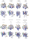
Comparison of SecYEG/Sec61 structures from various different studies. In each structure, SecY has been coloured in light blue, SecE in wheat and SecG in green, with the plug helix of SecY in red and the three SecY LG helices in dark blue (TMHs 2, 3 and 7; numbered accordingly in top left-hand panel). Where present, substrate helices are coloured in black (note that the substrate is not visible 4CG5, as the density was not assigned in the original structure).
Figure 5.
Figure 5.
Structures of SecA in a resting state (above, 1M6N [43]) and bound to SecYEG (below, 3DIN [31]), viewed from the cytoplasm (left) and the side (right). Key domains (NBD1, NBD2, PPXD and 2HF) are coloured and labelled; white regions comprise other domains not mentioned in the text. A pre-protein substrate has been modelled into SecA using known cross-linking sites [44] and energy minimized (marine spheres); note that closing of the PPXD (brown arrow) forms a clamp around the substrate. In the SecYEG-bound structures, SecYEG is shown as a mesh (although it is obscured by SecA in the left panel), with the 2HF of SecA inserted into the channel.
Figure 6.
Figure 6.
Proposed mechanisms of (a) Sec- and (b) Tat-mediated protein translocation. See §§3 and 4 for details.
Figure 7.
Figure 7.
One possible arrangement of TatA, TatB and TatC in complex. Model made using atomic models of individual components as follows: purple—solution NMR data for monomeric E. coli TatA, built into a ring and subjected to coarse grain and atomistic molecular dynamics simulation (PDB code 2LZS [102]); yellow—solution NMR structure of truncated (1–101) E. coli TatB monomer, with the flexible helices indicated with a yellow arrow (PDB code 2MI2 [101]); cyan—crystal structure of TatC from Aquifex aeolicus (PDB code 4B4A [27]). In each case, the models are aligned with respect to the membrane as per the original study. TatB and TatC have been aligned with respect to each other based on previous cross-linking data [27,104], and using the protein docking algorithm ZDOCK for feasible conformations [105]. Known cross-linking residues are shown as either green (TatB) or purple (TatC) spheres. The putative substrate-binding region is marked with an orange bar. Note that there are currently no known TatBC to TatA oligomer interaction sites.

Similar articles

Cited by

References

    1. Krogh A, Larsson B, von Heijne G, Sonnhammer E. 2001. Predicting transmembrane protein topology with a hidden Markov model: application to complete genomes. J. Mol. Biol. 305, 567–580. (10.1006/jmbi.2000.4315) - DOI - PubMed
    1. Song C, Kumar A, Saleh M. 2009. Bioinformatic comparison of bacterial secretomes. Genomics Proteomics Bioinform. 7, 37–46. (10.1016/S1672-0229(08)60031-5) - DOI - PMC - PubMed
    1. Blobel G, Dobberstein B. 1975. Transfer of proteins across membranes. I. Presence of proteolytically processed and unprocessed nascent immunoglobulin light chains on membrane-bound ribosomes of murine myeloma. J. Cell Biol. 67, 835–851. (10.1083/jcb.67.3.835) - DOI - PMC - PubMed
    1. Blobel G, Dobberstein B. 1975. Transfer of proteins across membranes. II. Reconstitution of functional rough microsomes from heterologous components. J. Cell Biol. 67, 852–862. (10.1083/jcb.67.3.852) - DOI - PMC - PubMed
    1. Görlich D, Rapoport TA. 1993. Protein translocation into proteoliposomes reconstituted from purified components of the endoplasmic reticulum membrane. Cell 75, 615–630. (10.1016/0092-8674(93)90483-7) - DOI - PubMed

Publication types

LinkOut - more resources